Case
Explore This Issue
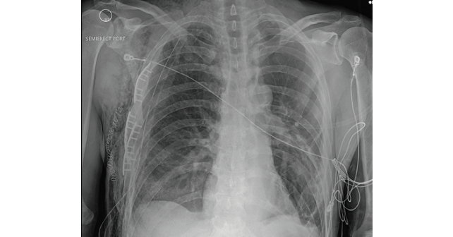
ACEP Now: Vol 42 – No 05 – May 2023FIGURE 1: Chest X-ray of multiple rib fractures (arrows). (Click to enlarge.)
A 58-year-old male with a history of alcohol abuse presented to the emergency department (ED) as a category 2 trauma for a fall with a reported flail chest. The patient had been drinking with friends when he was witnessed to trip and fall a distance of one step. His right chest wall struck a protuberance, initially reported to be the edge of a stair and later noted to be tree stump. EMS recognized a chest wall deformity with movement of the chest wall, and a splint was devised and taped around his chest for what was suspected to be a flail chest. The splint consisted of a folded blanket placed over the mobile segment and held in place with tape. The patient complained of right chest wall pain and shortness of breath that improved once splinted. He denied syncope, head trauma, or any other complaints. His vital signs were within normal limits except for a respiratory rate of 23 with a room air pulse oxygen in the upper 90s. Exam was notable for bilateral breath sounds though diminished on the right, with a mobile segment on the right lateral chest wall. The patient experienced significantly more discomfort with the splint removed and it was reapplied during his trauma bay evaluation. There were no open wounds and the remainder of his exam was unremarkable.

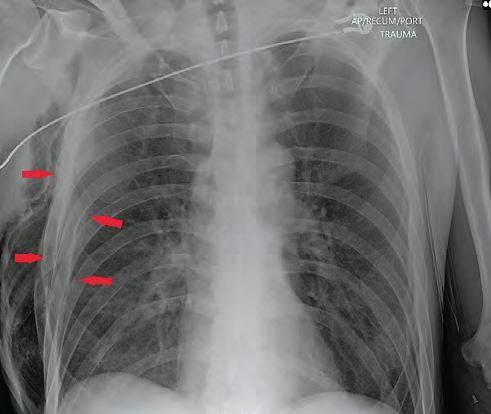
FIGURE 2: Axial chest CT of the chest wall defect (red arrow) and pneumothorax trauma (white arrows). (Click to enlarge.)
Trauma imaging was performed including bedside X-rays of the chest and pelvis. The chest X-ray showed multiple displaced right rib fractures and chest wall subcutaneous emphysema without definitive evidence of a pneumothorax, as well as right lower lobe atelectasis versus contusion (Figure 1). The patient was placed on oxygen for his dyspnea, administered pain medication, and was taken for computed tomography (CT). CTs of his head, spine, chest, abdomen, and pelvis were performed. A chest CT demonstrated multiple displaced right lateral rib fractures with direct communication between the pleural space and the soft tissue. There was evidence of a decompressed pneumothorax into the soft tissues of the right lateral chest wall with evidence of developing tension within the soft tissues. Extensive subcutaneous emphysema was dissecting from the chest disruption superiorly into the neck and inferiorly into the lower chest and abdominal wall (Figures 2 and 3).

FIGURE 3: Coronal CT lung window of subcutaneous air (red arrows), chest wall defect (white arrow).
Trauma labs were notable for a lactate of 3.0 mmol/L and a serum ethanol level of 160 mg/dL.
On re-evaluation, the chest wall movement was noted not to be following the paradoxical movement typical of flail segments. Instead, the flail segment was bulging outward with both inspiration and expiration (see Figure 4 video). The trauma team placed a pigtail catheter in the right chest cavity to decompress the pneumothorax and the patient was admitted to the surgical intensive care unit.
The patient continued to have an oxygen requirement and significant pain. On hospital day 2, he was taken to the operating room for surgical rib fixation. A chest tube was placed at that time. On postoperative day 5, the chest tube was removed, and he was discharged the following day (Figure 5).
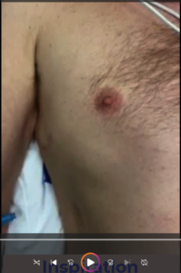
FIGURE 4: Click to play video of the patient breathing.
Discussion
Displaced rib fractures can injure lung tissue and cause a pneumothorax. In this case, the patient’s pneumothorax was decompressed into a large soft tissue defect in his chest wall. The extensive chest-wall disruption resulted in soft tissue emphysema that was bulging with respirations mimicking a flail chest. A flail chest is defined by multiple fractures in three or more consecutive ribs with paradoxical movement of the resulting chest wall segment. Flail chest can result in respiratory failure. Initial management includes analgesia and positive pressure ventilation to help stabilize the chest wall.1 Unlike the typical paradoxical chest wall movement seen with flail chest, the subcutaneous tissues in this case were inflating with both inspiration and expiration although this was not fully appreciated due to the significant discomfort the patient experienced when the splint was removed. The direct movement of air into the chest wall was improved with the placement of a pigtail catheter and ultimately treated with operative repair.

FIGURE 5: Chest X-ray OF post open reduction and interior fixation of rib fractures. (Click to enlarge.)
Traditionally, treatment of flail chest was aimed at associated injuries, especially pulmonary contusions, and supportive care. Definitive treatment with surgical stabilization has been gaining favor, with current literature suggesting decreased ICU stays and fewer complications, especially with patients under 60 years old when taken to the operating room within 72 hours of injury.2
Dr. Mitchell is an attending emergency physician in the department of emergency medicine at MetroHealth Medical Center in Cleveland, Ohio, as well as assistant professor at Case Western Reserve University School of Medicine in Cleveland, Ohio.
Dr. Baughman is staff physician in the department of radiology at MetroHealth Medical Center in Cleveland, Ohio, including Case Western Reserve University School of Medicine in Cleveland, Ohio.
Dr. Jones is professor of emergency medicine at Case Western Reserve University School of Medicine in Cleveland, Ohio. He is also system-wide clinical ultrasound co-chair at MetroHealth Medical Center in Cleveland, Ohio.
Dr. Effron is an attending physician in the department of emergency medicine at MetroHealth Medical Center, in Cleveland, Ohio, as well as associate professor of emergency medicine at Case Western Reserve University School of Medicine in Cleveland, Ohio.
References
- Mistry RN, Moore JE. Management of blunt thoracic trauma. BJA Education. 2022; 22(11):432-439.
- Sawyer E, Wullschleger M, Muller N, Muller M. Surgical rib fixation of multiple rib fractures and flail chest: a systematic review and meta-analysis. J Surg Res. 2022; 276:221-234.
Pages: 1 2 3 | Multi-Page


No Responses to “Case Report: EMS Says Flail Chest, But Is It?”