Logo

Log In Sign Up |  An official publication of: American College of Emergency Physicians
Navigation
  • Home
  • Multimedia
    • Podcasts
    • Videos
  • Clinical
    • Airway Managment
    • Case Reports
    • Critical Care
    • Guidelines
    • Imaging & Ultrasound
    • Pain & Palliative Care
    • Pediatrics
    • Resuscitation
    • Trauma & Injury
  • Resource Centers
    • mTBI Resource Center
  • Career
    • Practice Management
      • Benchmarking
      • Reimbursement & Coding
      • Care Team
      • Legal
      • Operations
      • Quality & Safety
    • Awards
    • Certification
    • Compensation
    • Early Career
    • Education
    • Leadership
    • Profiles
    • Retirement
    • Work-Life Balance
  • Columns
    • ACEP4U
    • Airway
    • Benchmarking
    • Brief19
    • By the Numbers
    • Coding Wizard
    • EM Cases
    • End of the Rainbow
    • Equity Equation
    • FACEPs in the Crowd
    • Forensic Facts
    • From the College
    • Images in EM
    • Kids Korner
    • Medicolegal Mind
    • Opinion
      • Break Room
      • New Spin
      • Pro-Con
    • Pearls From EM Literature
    • Policy Rx
    • Practice Changers
    • Problem Solvers
    • Residency Spotlight
    • Resident Voice
    • Skeptics’ Guide to Emergency Medicine
    • Sound Advice
    • Special OPs
    • Toxicology Q&A
    • WorldTravelERs
  • Resources
    • ACEP.org
    • ACEP Knowledge Quiz
    • Issue Archives
    • CME Now
    • Annual Scientific Assembly
      • ACEP14
      • ACEP15
      • ACEP16
      • ACEP17
      • ACEP18
      • ACEP19
    • Annals of Emergency Medicine
    • JACEP Open
    • Emergency Medicine Foundation
  • About
    • Our Mission
    • Medical Editor in Chief
    • Editorial Advisory Board
    • Awards
    • Authors
    • Article Submission
    • Contact Us
    • Advertise
    • Subscribe
    • Privacy Policy
    • Copyright Information

Case Report: EMS Says Flail Chest, But Is It?

By Alix Mitchell, MD; William Baughman, MD; Robert Jones, DO FACEP; David Effron, MD, FACEP | on May 4, 2023 | 0 Comment
Features
  • Tweet
  • Click to email a link to a friend (Opens in new window) Email
Print-Friendly Version

You Might Also Like
  • Case Report: Blunt Tracheal Injury Creates Difficult Airway
  • Case Report: Fitz-Hugh Curtis Syndrome in a Male with HIV
  • Case Report: An Endocrine Enigma
Explore This Issue
ACEP Now: Vol 42 – No 05 – May 2023

FIGURE 3: Coronal CT lung window of subcutaneous air (red arrows), chest wall defect (white arrow).

Trauma labs were notable for a lactate of 3.0 mmol/L and a serum ethanol level of 160 mg/dL.

On re-evaluation, the chest wall movement was noted not to be following the paradoxical movement typical of flail segments. Instead, the flail segment was bulging outward with both inspiration and expiration (see Figure 4 video). The trauma team placed a pigtail catheter in the right chest cavity to decompress the pneumothorax and the patient was admitted to the surgical intensive care unit.


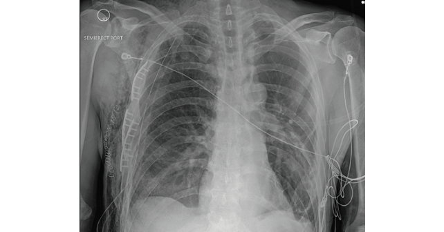
The patient continued to have an oxygen requirement and significant pain. On hospital day 2, he was taken to the operating room for surgical rib fixation. A chest tube was placed at that time. On postoperative day 5, the chest tube was removed, and he was discharged the following day (Figure 5).

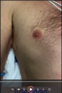
FIGURE 4: Click to play video of the patient breathing.

Discussion

Displaced rib fractures can injure lung tissue and cause a pneumothorax. In this case, the patient’s pneumothorax was decompressed into a large soft tissue defect in his chest wall. The extensive chest-wall disruption resulted in soft tissue emphysema that was bulging with respirations mimicking a flail chest. A flail chest is defined by multiple fractures in three or more consecutive ribs with paradoxical movement of the resulting chest wall segment. Flail chest can result in respiratory failure. Initial management includes analgesia and positive pressure ventilation to help stabilize the chest wall.1 Unlike the typical paradoxical chest wall movement seen with flail chest, the subcutaneous tissues in this case were inflating with both inspiration and expiration although this was not fully appreciated due to the significant discomfort the patient experienced when the splint was removed. The direct movement of air into the chest wall was improved with the placement of a pigtail catheter and ultimately treated with operative repair.

FIGURE 5: Chest X-ray OF post open reduction and interior fixation of rib fractures. (Click to enlarge.)

Traditionally, treatment of flail chest was aimed at associated injuries, especially pulmonary contusions, and supportive care. Definitive treatment with surgical stabilization has been gaining favor, with current literature suggesting decreased ICU stays and fewer complications, especially with patients under 60 years old when taken to the operating room within 72 hours of injury.2

Pages: 1 2 3 | Single Page

Topics: Case ReportsClinicalflail chestImaging & UltrasoundTrauma & Injury

Related

  • Non-Invasive Positive Pressure Ventilation in the Emergency Department

    October 1, 2025 - 0 Comment
  • Emergency Department Management of Prehospital Tourniquets

    October 1, 2025 - 0 Comment
  • ACEP’s October 2025 Poll: How Often Do You Read Your Own X-Rays?

    September 30, 2025 - 0 Comment

Current Issue

ACEP Now: November 2025

Download PDF

Read More

No Responses to “Case Report: EMS Says Flail Chest, But Is It?”

Leave a Reply Cancel Reply

Your email address will not be published. Required fields are marked *


*
*


Wiley
  • Home
  • About Us
  • Contact Us
  • Privacy
  • Terms of Use
  • Advertise
  • Cookie Preferences
Copyright © 2025 by John Wiley & Sons, Inc. All rights reserved, including rights for text and data mining and training of artificial technologies or similar technologies. ISSN 2333-2603