Although many cases of children presenting to the emergency department (ED) with bloody diarrhea are benign and self-limited, one potentially morbid diagnosis should always be considered: Shiga toxin-producing Escherichia coli (STEC). Although relatively rare, STEC infections carry the serious risk of hemolytic uremic syndrome (HUS), a leading cause of acute kidney injury in children. Shiga toxin 2 (Stx2) is specifically associated with a 15-20 percent risk for HUS in children younger than 5.1 In multiple case series studies, more than 50 percent of patients with STEC-related HUS required dialysis within one week.2 Emergency physicians must identify which children are at risk, initiate the right investigations, and arrange appropriate monitoring or admission to prevent severe morbidity or even mortality. By the end of this column, it’s my hope that you will gain an evidence-based approach to pediatric bloody diarrhea and be primed to recognize and manage STEC to improve outcomes.
Explore This Issue
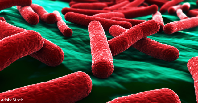
ACEP Now: August 2025 (Digital)When to Suspect STEC Infection
The evaluation of a child with bloody diarrhea begins, as always, with a thorough assessment of hemodynamic status and hydration. Fortunately, most children presenting with bloody diarrhea are hemodynamically stable and well-perfused, but subtle signs of dehydration or early shock should not be overlooked. In the unstable child, fluid resuscitation and empiric antibiotics are priorities. However, in most well-appearing, stable children, attention turns to determining the cause of the bleeding and assessing the risk for STEC infection.
Children with STEC infection frequently present with a short duration of illness, typically one to five days, marked by severe crampy abdominal pain and frequent small-volume bloody, mucous-like stools.3 Parents may report more than 15-20 bowel movements per day, and although fever may be present, it is often low-grade. The diarrhea tends to be of small volume, distinguishing it from the profuse watery diarrhea of cholera-like illnesses or large-volume bleeding from other causes. This pattern of frequent painful, bloody stools should raise suspicion for infectious colitis, with STEC as an important consideration. When should STEC infection be suspected? In children from endemic areas (which includes the northern states of the United States) who present with severe crampy abdominal pain, small frequent, mucous-like, bloody stools, low grade fever, and/or signs of microangiopathy such as petechiae and jaundice.4
Obtain a Stool Specimen or Rectal Swab
Although not all children who present to the ED with diarrhea require stool testing, those with bloody diarrhea should be considered for it. Clinicians should prioritize collecting a stool sample in the ED rather than sending the family home with a container for outpatient collection. Delaying specimen collection can prolong the time to diagnosis by a day or more, increasing the risk for missing the window for effective early intervention. Most laboratories in North America now use multiplex polymerase chain reaction (PCR) assays as the frontline diagnostic tool rather than culture. These panels typically test for STEC, Salmonella, Shigella, and Campylobacter, providing broad coverage of common bacterial pathogens.
When a child is unable to produce a stool sample in the ED, rectal swabs offer a valuable alternative, provided the microbiology lab accepts and processes such samples. This approach ensures testing begins immediately, avoiding delays inherent in outpatient collection. Early identification of STEC is crucial because it allows timely risk stratification and the opportunity for close follow-up or admission if needed.
Which Patients Require Blood Testing?
A common question in managing children with bloody diarrhea is whether bloodwork is necessary at the initial presentation. For the well-appearing, hemodynamically stable child with minimal bleeding, laboratory testing may offer little added value. However, several indications warrant baseline laboratory evaluation, including a complete blood count, creatinine, lactate dehydrogenase (LDH), and electrolytes. Bloodwork is indicated in children presenting with severe crampy abdominal pain and frequent small-volume bloody stools, as well as those with a history of recent travel, particularly if accompanied by fever. Close contact with a known STEC case, or residence in an area with a known outbreak, also increases pretest probability and supports baseline testing. Physical findings suggestive of microangiopathy, such as petechiae or jaundice, further justify early bloodwork.
Routine testing for parasites, particularly stool ova and parasites, is not recommended in patients with acute bloody diarrhea unless there is a relevant travel history, chronic symptoms, or other risk factors. Similarly, given the high carriage rates in asymptomatic infants and toddlers, testing for Clostridium difficile should not be routinely performed in young children without recent antibiotic exposure or hospitalization .5
Understanding STEC and Its Complications
Recognizing STEC is essential because of its potential progression to HUS. The pathophysiology of STEC hinges on its ability to produce Shiga toxin, a virulence factor capable of causing systemic endothelial injury. Once absorbed from the gut into the bloodstream, Shiga toxin binds to globotriaosylceramide (Gb3) receptors, which are most abundantly expressed in the renal glomeruli. The toxin’s cellular effects include inhibition of protein synthesis, endothelial damage, and microvascular thrombosis.2 This pathophysiologic cascade leads to the classic triad of HUS: microangiopathic hemolytic anemia, thrombocytopenia, and acute kidney injury.
Not all STEC strains carry the same risk of HUS. Some strains produce Shiga toxin 1 (Stx1), whereas others produce Stx2, or both. This distinction is clinically significant because Stx1-producing STEC carries a less than one percent risk for HUS, whereas Stx2-producing STEC is associated with a fifteen to twenty percent risk of HUS in children younger than five years. The serotype O157:H7, well known in medical literature, almost universally produces Stx2 and should be regarded as high risk.2
Risk Stratification of Patients with STEC Infection
Once a laboratory report confirms STEC, the next step is to determine whether the child is at high risk for progression to HUS. Risk stratification hinges on three key variables: the presence of bloody diarrhea, the duration of illness, and the toxin type if known.6 A child with STEC infection, bloody diarrhea, and less than 10 days of symptoms should be presumed high risk for HUS until proven otherwise. HUS develops a median of seven days after diarrhea onset. Children whose diarrhea has persisted for more than 10 days without signs of microangiopathy are generally considered low risk. If toxin typing identifies Stx2 or O157:H7, the child remains high risk regardless of symptom duration.6
Not all laboratories provide toxin typing or serotyping results within a clinically useful timeframe. Emergency physicians should be familiar with their laboratory’s reporting practices and if needed, communicate directly with the lab to clarify whether toxin typing is performed, and when results are expected. In many cases, toxin typing may require an additional 24 hours beyond initial PCR detection.
Management of High-Risk Patients
Management of a high-risk child centers on close monitoring and hydration. Literature suggests that intravascular volume depletion at the onset of HUS is associated with worse renal outcomes, including higher rates of dialysis and increased mortality.7 Preventing dehydration is a cornerstone of management. Children identified as high risk should undergo daily reassessment of hydration status, urine output monitoring, and serial laboratory testing, including complete blood count, creatinine, LDH (to identify hemolysis), and electrolytes.
The platelet count is the earliest laboratory marker of evolving HUS.2 A declining platelet count, even within the normal range, may indicate developing microangiopathy. For example, a drop from 400 to 275 x10⁹/L over 24 hours, although technically still normal, may represent early progression toward HUS in the context of STEC infection. Identifying such trends allows for early hospital admission and monitoring before the onset of overt renal failure or neurologic complications.
Children diagnosed with HUS require hospitalization, ideally in a center with pediatric nephrology expertise. Management includes careful attention to hydration, avoidance of nephrotoxic agents, monitoring for electrolyte disturbances and hypertension, and early recognition of neurologic complications such as seizures or altered mental status. Peritoneal dialysis is typically the preferred modality for renal replacement therapy in pediatric patients. Neurologic complications, including stroke and cerebral edema, are the most serious sequelae and the leading cause of mortality in HUS.8
Next time you are faced with a child with diarrhea, consider STEC infection, prioritize obtaining a stool specimen for PCR testing in the ED, stratify risk based on clinical features and laboratory findings, and monitor high-risk children closely for signs of evolving HUS. It is my hope that with the knowledge you gain from this column you will be better able to identify and manage STEC infections and help prevent progression to life-threatening renal and neurologic complications.
A special thanks to Dr. Stephen Freedman, the guest expert on the EM Cases podcast that inspired this column.

Dr. Helman is an emergency physician at North York General Hospital in Toronto. He is an assistant professor at the University of Toronto, Division of Emergency Medicine, and the education innovation lead at the Schwartz/Reisman Emergency Medicine Institute. He is the founder and host of the Emergency Medicine Cases podcast and website.
References
- Ylinen E, Salmenlinna S, Halkilahti J, et al. Hemolytic uremic syndrome caused by Shiga toxin-producing Escherichia coli in children: incidence, risk factors, and clinical outcome. Pediatr Nephrol. 2020;35(9):1749-1759.
- Freedman SB, van de Kar NCAJ, Tarr PI. Shiga toxin-producing Escherichia coli and the hemolytic-uremic syndrome. N Engl J Med. 2023;389(15):1402-1414.
- McKee RS, Tarr PI, Dietzen DJ, et al. Clinical and laboratory predictors of shiga toxin-producing escherichia coli infection in children with bloody diarrhea. J Pediatric Infect Dis Soc. 2018;7(3):e116-e122.
- Tack DM, Kisselburgh HM, Richardson LC, et al. Shiga toxin-producing Escherichia coli outbreaks in the United States, 2010-2017. Microorganisms. 2021;9(7):1529.
- Campbell CT, Poisson MO, Hand EO. An updated review of Clostridium difficile treatment in pediatrics. J Pediatr Pharmacol Ther. 2019;24(2):90-98.
- McKee RS, Schnadower D, Tarr PI, et al. Predicting hemolytic uremic syndrome and renal replacement therapy in Shiga Toxin-producing Escherichia coli-infected children. Clin Infect Dis. 2020;70(8):1643-1651.
- Scheiring J, Andreoli SP, Zimmerhackl LB. Treatment and outcome of Shiga-toxin-associated hemolytic uremic syndrome (HUS). Pediatr Nephrol. 2008;23(10):1749-1760.
- Mansour MA, Khalil DF, Hasham MA, et al. Hemolytic uremic syndrome with central nervous system manifestations, a case report and literature review. Radiol Case Rep. 2023;18(6):2268-2273.
Pages: 1 2 3 4 | Multi-Page


No Responses to “Pediatric Bloody Diarrhea: Recognition, Management of STEC Infection”