Ann Emerg Med. 2020 Feb 25. doi: 10.1016/j.annemergmed.2020.01.008. © ACEP
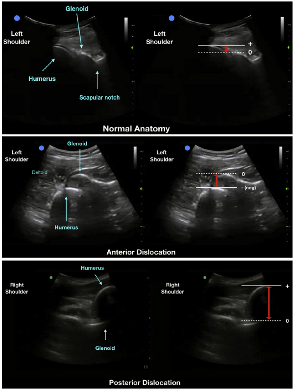
Ultrasonographic images of the shoulder girdle, using a curvilinear probe demonstrating the normal anatomy of the left glenohumeral joint (A), a left anterior dislocation of the shoulder with humeral head displaced anterior to the glenoid (B), and a right posterior shoulder dislocation with the humerus displaced posterior to the glenoid (C). The adjacent images correspond to the measurement of the glenohumeral distance, indicated by the red arrows.

No Responses to “Ann Emerg Med. 2020 Feb 25. doi: 10.1016/j.annemergmed.2020.01.008. © ACEP Ultrasonographic images of the shoulder girdle, using a curvilinear probe demonstrating the normal anatomy of the left glenohumeral joint (A), a left anterior dislocation of the shoulder with humeral head displaced anterior to the glenoid (B), and a right posterior shoulder dislocation with the humerus displaced posterior to the glenoid (C). The adjacent images correspond to the measurement of the glenohumeral distance, indicated by the red arrows.”