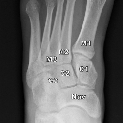
Figure 1: Normal AP weight-bearing radiograph of the left midfoot showing first metatarsal base (M1), second metatarsal (M2), third metatarsal (M3), medial cuneiform (C1), middle cuneiform (C2), lateral cuneiform (C3), and navicular bone (Nav). Note that there is less than 2 mm between C1 and M2 and between M1 and M2.
Figure 1: Normal AP weight-bearing radiograph of the left midfoot showing first metatarsal base (M1), second metatarsal (M2), third metatarsal (M3), medial cuneiform (C1), middle cuneiform (C2), lateral cuneiform (C3), and navicular bone (Nav). Note that there is less than 2 mm between C1 and M2 and between M1 and M2.
By Joseph Harrington
|
on December 13, 2017
|
0 Comment


No Responses to “Figure 1: Normal AP weight-bearing radiograph of the left midfoot showing first metatarsal base (M1), second metatarsal (M2), third metatarsal (M3), medial cuneiform (C1), middle cuneiform (C2), lateral cuneiform (C3), and navicular bone (Nav). Note that there is less than 2 mm between C1 and M2 and between M1 and M2.”