Logo

Log In Sign Up |  An official publication of: American College of Emergency Physicians
Navigation
  • Home
  • Multimedia
    • Podcasts
    • Videos
  • Clinical
    • Airway Managment
    • Case Reports
    • Critical Care
    • Guidelines
    • Imaging & Ultrasound
    • Pain & Palliative Care
    • Pediatrics
    • Resuscitation
    • Trauma & Injury
  • Resource Centers
    • mTBI Resource Center
  • Career
    • Practice Management
      • Benchmarking
      • Reimbursement & Coding
      • Care Team
      • Legal
      • Operations
      • Quality & Safety
    • Awards
    • Certification
    • Compensation
    • Early Career
    • Education
    • Leadership
    • Profiles
    • Retirement
    • Work-Life Balance
  • Columns
    • ACEP4U
    • Airway
    • Benchmarking
    • Brief19
    • By the Numbers
    • Coding Wizard
    • EM Cases
    • End of the Rainbow
    • Equity Equation
    • FACEPs in the Crowd
    • Forensic Facts
    • From the College
    • Images in EM
    • Kids Korner
    • Medicolegal Mind
    • Opinion
      • Break Room
      • New Spin
      • Pro-Con
    • Pearls From EM Literature
    • Policy Rx
    • Practice Changers
    • Problem Solvers
    • Residency Spotlight
    • Resident Voice
    • Skeptics’ Guide to Emergency Medicine
    • Sound Advice
    • Special OPs
    • Toxicology Q&A
    • WorldTravelERs
  • Resources
    • ACEP.org
    • ACEP Knowledge Quiz
    • Issue Archives
    • CME Now
    • Annual Scientific Assembly
      • ACEP14
      • ACEP15
      • ACEP16
      • ACEP17
      • ACEP18
      • ACEP19
    • Annals of Emergency Medicine
    • JACEP Open
    • Emergency Medicine Foundation
  • About
    • Our Mission
    • Medical Editor in Chief
    • Editorial Advisory Board
    • Awards
    • Authors
    • Article Submission
    • Contact Us
    • Advertise
    • Subscribe
    • Privacy Policy
    • Copyright Information

Identify and Treat Lisfranc Injuries

By Yenisleidy Paez Perez, DO | on January 4, 2018 | 0 Comment
CME Now Features
  • Tweet
  • Click to email a link to a friend (Opens in new window) Email
Print-Friendly Version
Figure 5: Normal three-column anatomy of Lisfranc complex. A(LEFT) shows the AP view. B(ABOVE) shows the oblique view. C(BELOW) shows the lateral view.

You Might Also Like
  • Tips for Catching Commonly Missed Ankle Injuries
  • How to Perform Ultrasound-Guided Forearm Nerve Blocks to Provide Non-Drug Pain Relief for Acute Injuries
  • Online Clinical Tool for Post–Bariatric Surgery Patients Could Improve Management
Explore This Issue
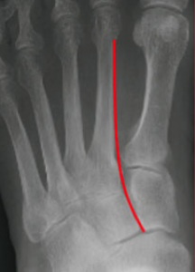
ACEP Now: Vol 36 – No 12 – December 2017

Figure 5: Normal three-column anatomy of Lisfranc complex. A shows the AP view.
Dr. Anton Helman/Emergency Medicine Cases

A(LEFT) shows the AP view.

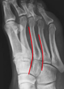
B shows the oblique view.

C shows the lateral view.

C shows the lateral view.
Diagnostic Imaging for the Emergency Physician. Elsevier 2011

X-Ray Findings Suspicious for Lisfranc Injuries

  • On the AP view, widening of >2 mm between the base of the first and second metatarsal indicates instability (See Figure 4).1
  • “Fleck sign” is pathognomonic for a Lisfranc injury. This is a small bony fragment avulsed from the second metatarsal base or medial cuneiform (see Figure 6).1

Management

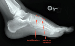
Stable dislocation/fracture injuries are defined as having less than 2 mm of displacement between the first metatarsal and medial cuneiform. These can be managed non-operatively with reduction and casting.5 The patient should be placed in a non-weight-bearing below-the-knee cast for six weeks and have outpatient orthopedic follow-up in two weeks.6

For unstable fractures and dislocations, immediate orthopedic consultation is needed for surgical intervention with internal fixation.5 After surgery, immobilization and non-weight-bearing status is recommend for eight to 12 weeks.7 The screws may then be removed at 12 weeks.7 Full weight-bearing is typically not permitted until all hardware is removed.

Complete the CME activity.


Dr. Paez PerezDr. Paez Perez is an emergency medicine resident at St. Joseph’s Regional Medical Center in Paterson, New Jersey.

References

  1. Siddiqui NA, Galizia MS, Almusa E, et al. Evaluation of the tarsometatarsal joint using conventional radiography, CT, and MR imaging. Radiographics. 2014;34(2):514-531.
  2. Lau S, Bozin M, Thillainadesan T. Lisfranc fracture dislocation: a review of a commonly missed injury of the midfoot. Emerg Med J. 2017;34(1):52-56.
  3. Caswell F, Brown C. Identifying foot fractures and dislocations. Emerg Nurse. 2014;22(6):30-34.
  4. Englanoff G, Anglin D, Hutson HR. Lisfranc fracture-dislocation: a frequently missed diagnosis in the emergency department. Ann Emerg Med. 1995;26(2):229-233.
  5. Anderson RB, Hunt KJ, McCormick JJ. Management of common sports-related injuries about the foot and ankle. J Am Acad Orthop Surg. 2010;18(9):546-556.
  6. Buzzard BM, Briggs PJ. Surgical management of acute tarsometatarsal fracture dislocation in the adult. Clin Orthop Relat Res. 1998;(353):125-133.
  7. Harwood MI, Raikin SM. A Lisfranc fracture-dislocation in a football player. J Am Board Fam Pract. 2003;16(1):69-72.

Pages: 1 2 3 | Single Page

Topics: CMECME NowEmergency DepartmentEmergency MedicineEmergency PhysiciansfractureimagingPatient CareTrauma and InjuryX-Ray

Related

  • Patterns of Injury in Elder Abuse

    November 6, 2025 - 0 Comment
  • Poll: Emergency Physicians Read Their Own X-Rays More Often Than Not

    November 6, 2025 - 0 Comment
  • Let Core Values Help Guide Patient Care

    November 5, 2025 - 0 Comment

Current Issue

ACEP Now: November 2025

Download PDF

Read More

No Responses to “Identify and Treat Lisfranc Injuries”

Leave a Reply Cancel Reply

Your email address will not be published. Required fields are marked *


*
*


Wiley
  • Home
  • About Us
  • Contact Us
  • Privacy
  • Terms of Use
  • Advertise
  • Cookie Preferences
Copyright © 2025 by John Wiley & Sons, Inc. All rights reserved, including rights for text and data mining and training of artificial technologies or similar technologies. ISSN 2333-2603