Figure 1
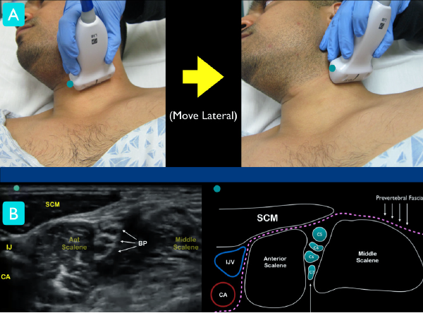
Figure 1A: Classic technique for locating the interscalene brachial plexus. At the level of the larynx, slide lateral until the ultrasound landmarks are noted
Figure 1B: Just under the sternocleidomastoid muscle (SCM), locate the anterior and middle scalene muscles. The interscalene groove will contain the nerve roots of the brachial plexus. Note the internal jugular vein (IJV) and carotid artery (CA) on the medial aspect of the anterior scalene muscle.

No Responses to “Figure 1 Figure 1A: Classic technique for locating the interscalene brachial plexus. At the level of the larynx, slide lateral until the ultrasound landmarks are noted Figure 1B: Just under the sternocleidomastoid muscle (SCM), locate the anterior and middle scalene muscles. The interscalene groove will contain the nerve roots of the brachial plexus. Note the internal jugular vein (IJV) and carotid artery (CA) on the medial aspect of the anterior scalene muscle.”