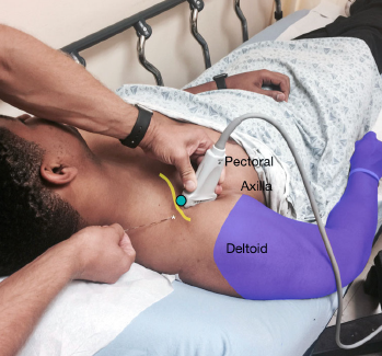
Figure 2: Stand above the patient with the ultrasound screen in clear view. Note the transducer is located in the deltopectoral groove with the needle entering above the clavicle.
Figure 2: Stand above the patient with the ultrasound screen in clear view. Note the transducer is located in the deltopectoral groove with the needle entering above the clavicle.

No Responses to “Figure 2: Stand above the patient with the ultrasound screen in clear view. Note the transducer is located in the deltopectoral groove with the needle entering above the clavicle.”