Lacerations to the hand are common injuries that present to emergency departments, both in academic Level 1 trauma centers as well as community and rural settings. Lacerations across the dorsal surface of the hand and fingers often injure the extensor tendons of the fingers, leading to difficulty or inability to extend the joints of the finger.
Explore This Issue
ACEP Now: March 2026The repair of these tendons is essential to preserving function of the hand and can be performed by a hand surgeon, but it is a relatively simple procedure, and in a resource-limited setting, can be performed by an emergency physician.
Anatomy of Extensor Tendons
For a brief review of the anatomy and physiology of the extensor tendons, the tendons from muscles of the forearm meet the tendons of the hand in a sheet at the base of each finger (near the knuckle). The extensor digitorus communis (EDC) muscle of the forearm supplies tendons that work to extend the index, middle, ring, and small fingers. The index finger has two tendons (EDC and extensor indices proprius). The extensor digiti minimi muscle (EDM), in addition to the EDC, works in conjunction to extend the fifth digit. The extensor pollicis brevis and longus muscles supply the tendons for the thumb. The sheet of tendinous material gives off a central band that extends the middle joint of the finger, and two separate lateral bands that converge together past the joint and function to extend the distal interphalangeal (DIP) joint. Unlike flexor tendons, extensors usually do not retract significantly because of connections called juncturae tendinae.
Diagnosis and Repair
To diagnose an extensor tendon injury, the patient will have pain with, or an inability to extend, one of the joints of the finger or finger drooping. Traumatic etiology of extensor tendon injury involves penetrating or blunt trauma to or with extension to the dorsal surface of the wrist, hand, or finger.
Once diagnosed, repair can be attempted. To prepare for the procedure, use a blood pressure cuff around the forearm and inflate it to 50 mmHg above systolic pressure to control bleeding. You can achieve anesthesia with digital ring block, local anesthetic, or systemic analgesia or moderate sedation, as needed. Liberally irrigate site of injury with 500 mL to 1 L of normal saline and apply sterile drapes around site of procedure.
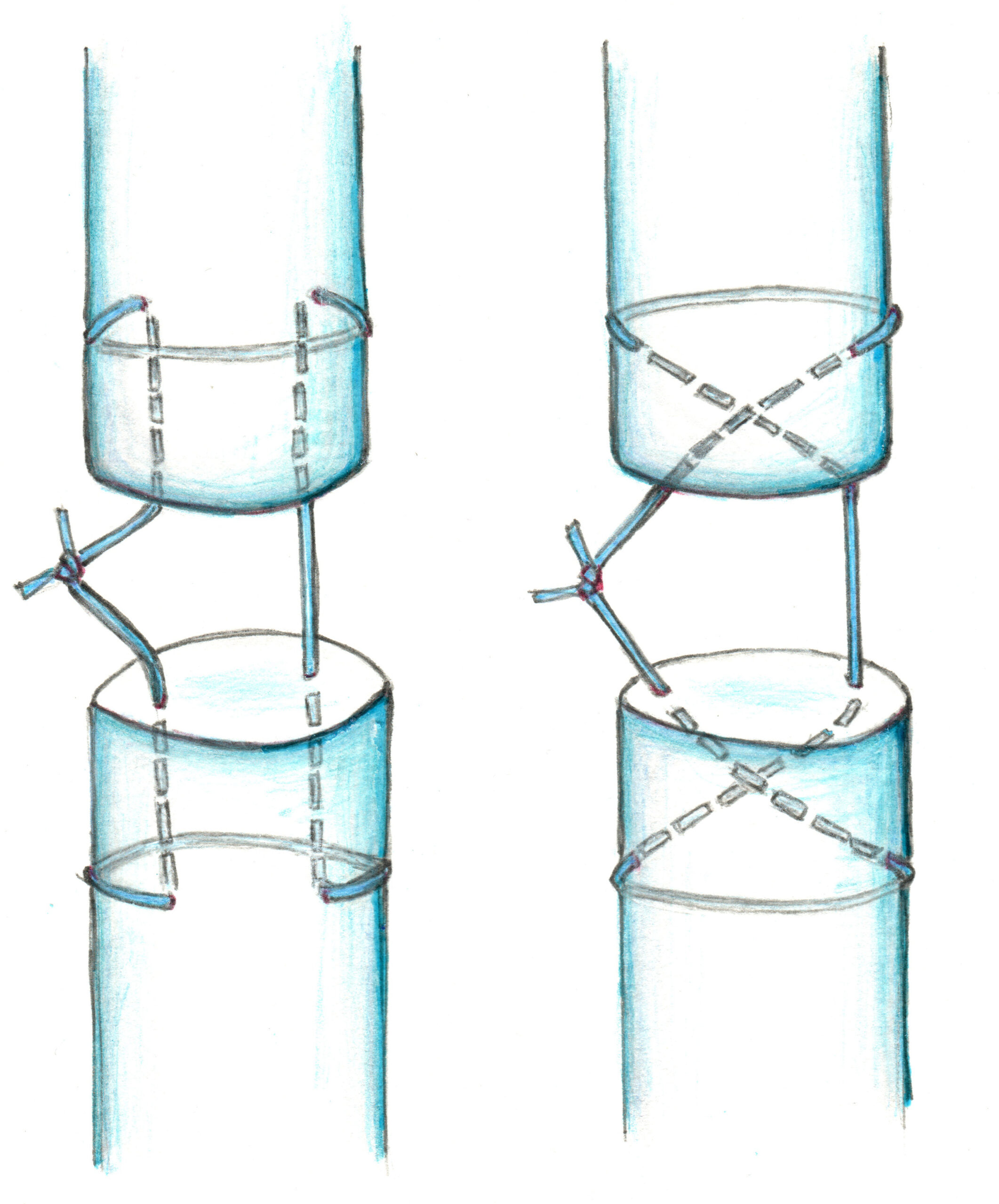
– FIGURE 1A (LEFT): Modified Kessler stitch
– FIGURE 1B (RIGHT): Modified Bunnell stitch
(Click to enlarge. ILLUSTRATIONS: KRISTINA K. ROBINSON, MD)
The repair is performed by extending the site of injury to visualize both ends of the severed tendon and using sutures to join the cut ends together. For injuries to the extensor tendon when it travels through the hand or proximal finger, the Modified Kessler stitch or Modified Bunnell stitch can be used (see Figure 1).
Figure 1A – Modified Kessler stitch: Insert the needle perpendicular to the proximal severed surface of tendon along the radial side (closest towards thumb), then exit tendon laterally and wrap the string around the tendon. Insert needle into ulnar side of tendon at approximately the same distance from the lacerated edge where the needle pierced the radial side, then exit the tendon perpendicularly through the severed end of tendon at a 90-degree angle. Now, join the proximal tendon with the distal tendon by entering the distal tendon severed surface on ulnar side, exit out the tendon laterally, wrap around the tendon, and reenter the distal tendon on radial side, exiting through severed surface on the radial half. Tighten the string and tie a knot that will be buried between the two segments of tendon.
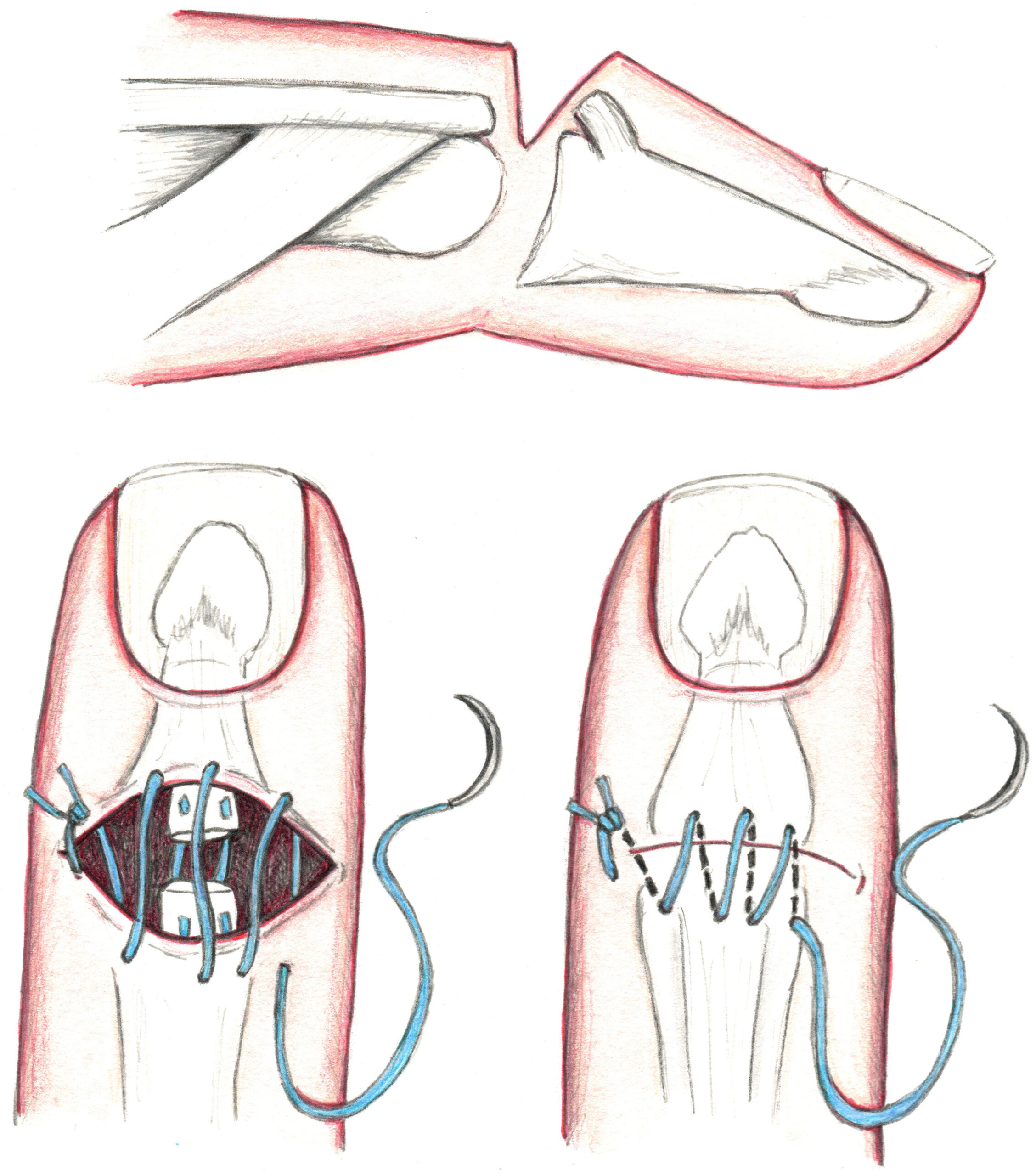
FIGURE 2: Roll suture. (Click to enlarge.)
Figure 1B – Modified Bunnell stitch: Similar to the Kessler stitch in that the needle is inserted into proximal severed surface of tendon towards the radial side, but instead of exiting the tendon on that same side, the needle is guided diagonally towards ulnar side to exit the tendon. Wrap the suture around tendon and reenter tendon on radial side at same distance from severed surface that you exited on the opposite side. Travel diagonally to exit out severed surface towards the ulnar surface. Repeat the process on the distal end of the tendon except by starting at the ulnar surface and ending by exiting radial surface. Join the two fragments of tendon together and tighten the string, tying a buried knot.
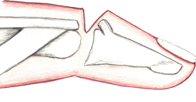
Injury to the tendon at the distal finger near the DIP joint should be repaired by dermatotenodesis (see Figure 2). This is performed by anchoring a simple suture on one end of the skin incision, leaving the tail of the suture long enough to perform the rest of the repair. Then perform running sutures until you reach the area where the tendon is located, and upon reentering skin on distal end of the laceration take a bite into superior surface of distal tendon on its lateral side (⅓ through diameter), exiting out through severed surface of distal tendon. Enter the proximal tendon through the lateral aspect of the severed surface, and exit out through the superior surface, also grabbing overlying skin. Repeat this technique by reentering the skin that is overlying medial ⅓ tendon and grabbing distal and proximal tendon fragments, exiting out through the skin. Place a single suture at the medial end of the incision to anchor the skin.
For all repairs, apply a volar splint to keep the affected tendon and joints held in extension for at least six weeks, making sure that the dressing isn’t too tight and the fingers are well perfused. Refer the patient to a hand surgeon or orthopedic surgeon for follow up. The splint pictured below can be used for injuries to the tendon at the distal finger involving DIP joint (repaired using dermatotenodesis).
Common complications after extensor tendon repair include infection, adhesions resulting in decreased mobility of hand, mallet deformity or swan neck deformity of finger, or ulceration to skin from splint application.

Dr. Dooley is an Assistant Professor of Emergency Medicine at Baylor College of Medicine in Houston, Texas.
 Dr. Smith is an Assistant Professor of Plastic Surgery at the University of Texas Medical Branch.
References
- The British Society for Surgery of the Hand. Extensor tendon injury. 2017. Accessible at https://www.bssh.ac.uk/patients/conditions/27/extensor_tendon_injury.
- Baecher, NB. Extensor Tendon Repair Technique. MedScape. 2023. Accessible at https://emedicine.medscape.com/article/109111-technique?scode=msp&st=fpf&socialSite=google&form=login&errorCode=invalidCombination®=1&icd=login_success_email_match_norm.
- The American Society for Surgery of the Hand. Handcare: The Upper Extremity Expert. 2025. Accessible at: https://www.assh.org/handcare/safety/tendons#Finger%20Extensor.


No Responses to “Extensor Tendon Repair by Emergency Physicians”