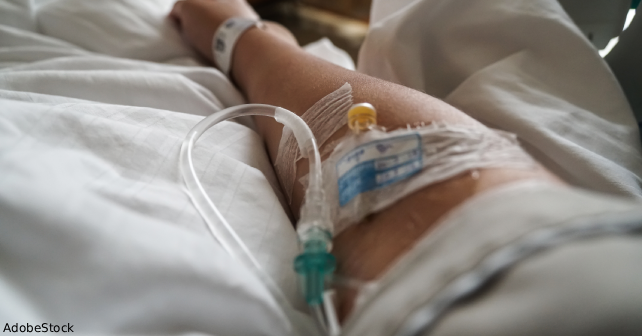
A 39-year-old man deployed in Afghanistan presented to a field medical station with acute gastroenteritis. He received a peripheral intravenous catheter (PIVC) in the right cephalic vein for fluid resuscitation, receiving 2 L over two hours. The PIVC was removed after fluid administration. Over the next few days, the patient noticed mild pain and soreness in the antecubital fossa, which was treated with acetaminophen and ibuprofen.
Explore This Issue
ACEP Now: October 2025 (Digital)Five days after IV insertion, the patient developed increased pain and noted a palpable cord extending from mid-forearm to biceps. Bedside emergency department ultrasound revealed thrombosis extending from the cephalic vein in the mid-forearm to the axillary vein, confirming upper extremity deep vein thrombosis (UE-DVT). He was started on low molecular weight heparin (LMWH) and aspirin. The next morning, he was evacuated to a higher echelon of care for imaging.
Upon arrival to a facility with a higher level of care, computed tomography of the right-sided venous system confirmed DVT extending from the cephalic vein to the subclavian vein with multiple right-sided segmental pulmonary embolism (PE). No imaging of the left-sided lung was obtained. He was started on rivaroxaban and was evacuated out of Afghanistan.
Notably, the patient reported a history of superficial thrombophlebitis in the same vein in 2014 after a peripheral IV insertion, which was treated with aspirin and warm compresses. No central lines or known malignancy were present. Hypercoagulability workup was deferred because of the provoked nature of the event.
Discussion
PIVC placement is the most frequently performed invasive medical procedure worldwide. Although the majority of complications are local and self-limited, serious vascular sequelae including DVT and PE are rarely reported, especially when arising from the upper extremity.1 Upper extremity DVT comprises only 5-10 percent of all DVTs but carries a risk of embolic complications.1 The transition from superficial venous thrombosis (SVT) to DVT and PE is underrecognized, particularly in patients without central venous catheters or malignancy.2 This case highlights a rare but important chain of events stemming from PIVC use in a deployed military environment, complicated by prior venous disease and acute gastroenteritis.
Though PIVC insertion is often considered low risk, this case demonstrates its potential to initiate a severe thrombotic cascade. SVT is usually self-limited, but studies show that progression to DVT can occur in up to 20-30 percent of patients, particularly when the thrombus is near a junction with deep veins.3 In this case, involvement of the cephalic vein—draining directly into the axillary-subclavian system—likely facilitated propagation.
Upper extremity DVT has traditionally been associated with central venous catheters, pacemakers, thoracic outlet syndrome, or malignancy. However, emerging data show that even peripheral interventions may cause endothelial injury sufficient to provoke thrombus formation, particularly when compounded by systemic risk factors such as infection, dehydration, and prior venous damage.2,4
The rate of PE occurrence in patients with UE-DVT is rarely reported. Additionally, as this patient had prior thrombophlebitis in the same location, it is plausible that local scarring and valvular dysfunction promoted stasis and increased the thrombogenic potential.3
Environmental stressors in the field like dehydration, high-altitude physiology, and limited diagnostic resources may amplify the risk for thrombosis.5 The gastroenteritis episode may have induced a transient hypercoagulable state through hemoconcentration and inflammatory cytokine release.6
Early identification of SVT, particularly in high-risk individuals, should prompt duplex ultrasonography to evaluate for contiguous DVT. Management of UE-DVT mirrors that of lower extremity DVT, including prompt initiation of anticoagulation. Guidelines from the American College of Chest Physicians support anticoagulation for symptomatic UE-DVT for at least three months.5
Conclusion
This case underscores the potential for a seemingly routine procedure like PIVC insertion to result in serious thromboembolic complications, particularly within the unique context of a deployed military environment. The progression from superficial thrombophlebitis to UE-DVT and ultimately to PE highlights a continuum of vascular injury that may be underrecognized, especially in patients lacking classic risk factors such as malignancy or central venous catheters.
The austere conditions of deployment, limited diagnostic capabilities, environmental stressors like dehydration and high altitude, and delayed access to advanced care can all exacerbate thrombotic risk. This patient’s prior history of venous injury, combined with a transient hypercoagulable state because of acute gastroenteritis, created a confluence of factors that contributed to thrombus formation and embolic progression. Emergency and military medicine physicians must be particularly vigilant for such complications in deployed settings, where early signs of vascular inflammation may be easily overlooked or attributed to benign causes.
This case emphasizes the importance of thorough assessment of intravenous access sites, particularly in patients with known prior vein injury or systemic stressors. Physicians should maintain a high index of suspicion for thrombotic events in patients presenting with pain, swelling, or palpable cords after PIVC insertion. When available, point-of-care ultrasound can facilitate early diagnosis and guide prompt anticoagulation, potentially averting life-threatening sequelae such as PE.
Although existing guidelines such as those from the American College of Chest Physicians support anticoagulation for symptomatic UE-DVT, they often focus on patients with central venous catheters or malignancy.5 As the operational and civilian medical communities continue to report similar cases, there is a need to better characterize thrombotic risk associated with peripheral IVs, especially in high-risk environments like combat zones.
In conclusion, this case serves as a cautionary reminder that no medical procedure is truly benign. It calls for increased awareness, surveillance, and preventive strategies surrounding even routine interventions like PIVC placement, especially in emergency and deployed military settings. Proactive recognition and timely management of thrombotic complications can significantly improve outcomes for service members and civilians alike.
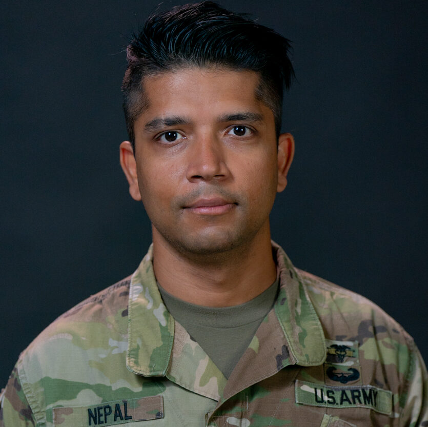 Dr. Nepal is a third-year emergency medicine resident at Madigan Army Medical Center. He completed his medical education at the Uniformed Services University of the Health Sciences. Dr. Nepal was born and raised in Nepal before moving to the U.S. for college at Kutztown University in Pennsylvania. Prior to medical school, he served as an enlisted soldier in the U.S. Army, a journey that continues to shape his dedication to service and patient care with humility.
References
- Valeriani E, Di Nisio M, Porceddu E, et al. Anticoagulant treatment for upper extremity deep vein thrombosis: a systematic review and meta-analysis. J Thromb Haemost. 2022;20(3):661-670.
- Duan Y, Wang GL, Guo X, et al. Acute pulmonary embolism originating from upper limb venous thrombosis: two case reports. World J Clin Cases. 2022;10(21):7445–7450.
- Lebon M, Ancedy Y, Boccara F, et al. Foreign body causing superficial venous thrombosis and subsequent pulmonary embolism: a case report. Eur Heart J Case Rep. 2018;2(4):yty125.
- Chen AW, Oraii Yazdani K, Candilio L. Upper limb deep vein thrombosis: a case report of an increasingly common condition. J Tehran Heart Cent. 2018;13(2):73–75.
- Kearon C, Akl EA, Ornelas J, et al. Antithrombotic therapy for VTE disease: CHEST guideline and expert panel report. Chest. 2016;149(2):315–352.
- Li Y, Wang Z, Sang X. Subclavian vein thrombosis and fatal pulmonary embolism after proximal humerus fracture surgery. J Orthop Surg Res. 2019;14:76.
Pages: 1 2 3 | Multi-Page


One Response to “Case Report: Rare Pulmonary Embolism After Routine PIVC Insertion”
September 28, 2025
Scott MankowitzInteresting story. Please stop creating new abbreviations and placing them in the title of the article.