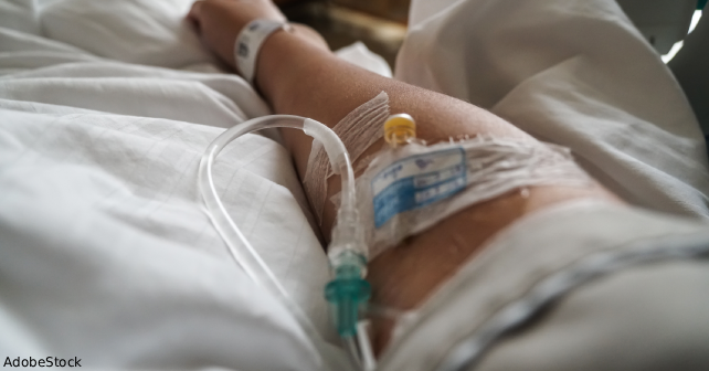
A 39-year-old man deployed in Afghanistan presented to a field medical station with acute gastroenteritis. He received a peripheral intravenous catheter (PIVC) in the right cephalic vein for fluid resuscitation, receiving 2 L over two hours. The PIVC was removed after fluid administration. Over the next few days, the patient noticed mild pain and soreness in the antecubital fossa, which was treated with acetaminophen and ibuprofen.
Explore This Issue
ACEP Now: October 2025 (Digital)Five days after IV insertion, the patient developed increased pain and noted a palpable cord extending from mid-forearm to biceps. Bedside emergency department ultrasound revealed thrombosis extending from the cephalic vein in the mid-forearm to the axillary vein, confirming upper extremity deep vein thrombosis (UE-DVT). He was started on low molecular weight heparin (LMWH) and aspirin. The next morning, he was evacuated to a higher echelon of care for imaging.
Upon arrival to a facility with a higher level of care, computed tomography of the right-sided venous system confirmed DVT extending from the cephalic vein to the subclavian vein with multiple right-sided segmental pulmonary embolism (PE). No imaging of the left-sided lung was obtained. He was started on rivaroxaban and was evacuated out of Afghanistan.
Notably, the patient reported a history of superficial thrombophlebitis in the same vein in 2014 after a peripheral IV insertion, which was treated with aspirin and warm compresses. No central lines or known malignancy were present. Hypercoagulability workup was deferred because of the provoked nature of the event.
Discussion
PIVC placement is the most frequently performed invasive medical procedure worldwide. Although the majority of complications are local and self-limited, serious vascular sequelae including DVT and PE are rarely reported, especially when arising from the upper extremity.1 Upper extremity DVT comprises only 5-10 percent of all DVTs but carries a risk of embolic complications.1 The transition from superficial venous thrombosis (SVT) to DVT and PE is underrecognized, particularly in patients without central venous catheters or malignancy.2 This case highlights a rare but important chain of events stemming from PIVC use in a deployed military environment, complicated by prior venous disease and acute gastroenteritis.
Though PIVC insertion is often considered low risk, this case demonstrates its potential to initiate a severe thrombotic cascade. SVT is usually self-limited, but studies show that progression to DVT can occur in up to 20-30 percent of patients, particularly when the thrombus is near a junction with deep veins.3 In this case, involvement of the cephalic vein—draining directly into the axillary-subclavian system—likely facilitated propagation.
Pages: 1 2 3 | Single Page


One Response to “Case Report: Rare Pulmonary Embolism After Routine PIVC Insertion”
September 28, 2025
Scott MankowitzInteresting story. Please stop creating new abbreviations and placing them in the title of the article.