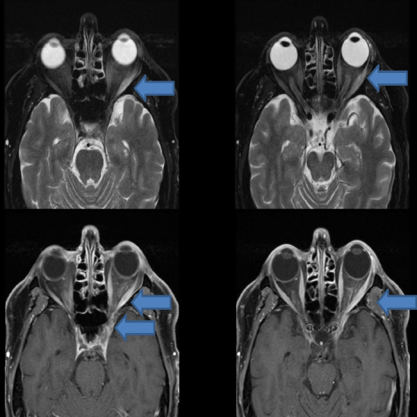
The patient’s MRI revealed an increased T2 signal with abnormal enhancement within the left lateral rectus muscle and intraconal fat extending into the orbital apex and left cavernous sinus.
The patient’s MRI revealed an increased T2 signal with abnormal enhancement within the left lateral rectus muscle and intraconal fat extending into the orbital apex and left cavernous sinus.

No Responses to “The patient’s MRI revealed an increased T2 signal with abnormal enhancement within the left lateral rectus muscle and intraconal fat extending into the orbital apex and left cavernous sinus.”