A 44 year-old male with unknown past medical history came by emergency medical services (EMS) to the emergency department (ED) for an electrical injury and fall from a high voltage electrical pole. Per EMS, the patient was found at the bottom of a high voltage line with diffuse burns and amputation of his left forearm. The patient was Glasgow Coma Scale (GCS) 15 on scene and complaining of back pain. He was intubated by EMS due to the extent of his injuries. His vitals were stable and endotracheal tube confirmed with end capnography and chest radiography.
Explore This Issue
ACEP Now: Vol 43 – No 03 – March 2024
FIGURE 1: Electrical burns of the patient’s bilateral feet. (Click to enlarge.)
The patient had a small left pneumo-thorax. A left thoracostomy, foley, nasogastric tube, and central venous catheter were placed prior to computed tomography (CT) imaging. The patient’s initial troponin was 17.78 ng/mL, creatine kinase greater than 60,000 U/L, glucose of 53 mmol/L, creatinine of 1.88 mg/dL and potassium of 5.7 mEq/L. He had estimated 45 percent total body surface area burns and received lactated Ringer’s infusion per Burns Rule of TENS (estimating to the nearest 10 percent TBSA and multiplying by 10 for the initial mL/hr; 40-80 kg adults) cefazolin, D50, warming, calcium gluconate, and tetanus.

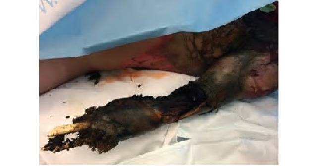
FIGURE 2: Traumatic amputation and electrical burn of the patient’s left arm. (Click to enlarge.)
Electrical injuries—excluding lightning injuries—account for roughly 10,000 nonfatal shock incidents a year and 500 deaths a year. While uncommon, electrical injuries disproportionately account for five percent of burn admissions in the U.S. and five percent workplace-related deaths.1-3 It’s important to understand the nuances of electrical injuries in order to identify hidden injuries and appropriately treat them.
Quick Review of the Physics

FIGURE 3: Electrical
burns of the patient’s leg with proximal tibia intraosseous line. (Click to enlarge.)
A brief review of the physics of electricity can help with clinical understanding. Electricity, in the form of electrons, travel down a gradient from high to low potential. The difference of the potential is the voltage (V). The “amount” of electrons in a timeframe down this gradient is the current. Resistance is the impedance of these electrons by the material and dissipates energy as heat. Our bodies have varying amounts of resistance—the higher the fluid and electrolyte content, the less resistance there is. Our skin is the barrier to prevent electricity from traveling into deeper tissues but varies in its resistance. Wet, thin skin of a child who just got out of a pool will allow electricity to pass into deeper tissues, which can lead to unseen internal burns. Thick, dry, and calloused skin of a construction worker can have as much as 100 times more resistance than the previous example.4 This may lead to more heat dissipated at the skin with impressive burns to skin, but less transmission of electricity to deeper tissues. Current can be alternating current (AC) or direct current (DC) with AC typically more dangerous as it is more likely to cause tetanic contractions and increase contact time with the electrical source. “High voltage” is defined by texts as 600 V and 1,000 V.2,3,5 Except for laundry or electrical car outlets (240 V AC), all U.S. household outlets are rated at 120 V AC. This means most household injuries are low voltage, with high voltage injuries happening in industrial settings or associated with power lines.

FIGURE 4: Computed tomography of abdomen and pelvis with edematous bowel. (Click to enlarge.)
The Unstable, Electrical Injury Patient
In evaluating a patient with electrical injuries, an approach to the stability of the patient should always take precedence. An unstable patient who is altered or with tenuous vital signs should consider trauma and cardiac causes. 10 percent of high-voltage electrical injuries will have an associated, significant traumatic injury.4,5 High voltage injuries can throw a victim from the electrical source, lead to falls, and cause forceful tetany with spinal hyperextension injuries or joint dislocations. Thus, unstable patients should be stabilized based on a physician’s trauma expertise; whether through Advanced Trauma Life Support, Trauma Combat Casualty Care, or one’s own trauma assessment. In addition, electrical injuries can affect cardiac conduction, leading to dysrhythmias with anything from atrial fibrillation to ventricular fibrillation.1,4-5 Thus, an electrocardiogram (ECG) and cardiac monitoring should be performed for unstable, electrical injury patients.
Three Injury Patterns: Trauma, Burns, and Electroporation Injuries
Either after stabilization or in a stable electrical injury patient, it may be helpful to categorize and assess for three different injury patterns of trauma, burns, and electroporation injuries.
Burns in electrical injuries can vary from superficial to full thickness based on skin resistance. For high voltage electrical injuries, a high clinical suspicion and thorough evaluation should be performed for any internal burns, even without significant skin findings. Bone has high resistance, leading to periosteal and surrounding myonecrosis. Cardiac muscle can be burned, leading to significant rises in troponin. However, this is not likely due to occlusive myocardial infarction requiring catheterization, but rather direct cardiac muscle damage or vasospasm.4,6 Internal viscera, such as bowel, can be burned, but is less likely given its high relative electrolyte and fluid content. Lastly, vessels can be damaged by internal burns, which may lead to poor perfusion and delayed complications as thrombosis or third spacing. Treatment for burns should focus on fluid resuscitation as appropriate based on your institution’s burn protocol per Brooke, Parkland, Rule of 10s etc. In addition, subacute injuries as compartment syndrome and rhabdomyolysis should be considered with creatine kinase, urinalysis, electrolyte panel, and monitoring urine output.
Electroporation injuries are caused by inappropriate membrane depolarization to electrically sensitive tissues such as the cardiac electrical conduction system or the nervous system. Dysrhythmias can present as syncope and/or chest pain, and albeit rare, have presented up to 12 hours after injury in low voltage injuries.7 Thus, an ECG is recommended for all electrical injuries. Syncope can also occur due to disruption of central nervous tissue. Other electrically sensitive nervous tissue can be damaged, presenting with paresthesias, weakness, and cognitive dysfunction or mood alterations. These can be delayed, up to weeks later, so it’s important to educate patients on the importance of follow-up with burn care well-versed with electrical injuries or neurology if they have or develop these symptoms.8,9
Disposition
Disposition depends on the patient’s clinical status and voltage exposure. All high voltage injuries should be transferred and treated at a regional burn center.10 Cardiac monitoring and observation of at least eight hours should be considered for a patient with an electrical injury and isolated syncope.5 Otherwise, if a patient’s ECG is normal, a low voltage exposure, and the clinical examination is without any significant trauma, burns, or electroporation injuries, the patient may be safely discharged with follow-up as warranted.
In our case, the patient had a CT of the head, cervical spine, and chest, abdomen, and pelvis with intravenous contrast demonstrated a C2, C7 compression fracture with significant bowel edema (Figure 4), and was taken to the operating room for left arm disarticulation, left below knee amputation and right above knee amputation. His ECG was unremarkable. In the operating room, there was minimal urinary output and the bladder pressures were 35 mmHg under sedation and analgesia. Subsequently, a decompressive laparotomy was performed for abdominal compartment syndrome. He underwent continuous renal replacement therapy (CRRT) with complications of acute respiratory distress syndrome and need for left scapular disarticulation, and transitioned to comfort care by his next of kin on hospital day five.
 Dr. Koo is faculty and an emergency physician at MedStar Washington Hospital Center in Washington, D.C., and St. Mary’s Hospital in Leonardtown, Maryland.
 Dr. McCollum is the director of teaching and learning in the department of emergency medicine at Augusta University in Augusta, Georgia.
References
- Zemaitis MR, Foris LA, Lopez RA, Huecker MR. Electrical Injuries. 2023 Jul 17. In: StatPearls [Internet]. Treasure Island (FL): StatPearls Publishing; 2023 Jan– PMID: 28846317.
- Tintinalli JE, Stapczynski J, Ma O, et al. eds. Tintinalli’s Emergency Medicine: A Comprehensive Study Guide, 8e. McGraw Hill; 2016. Accessed October 21, 2023.
- Electrical Safety Foundation International. ESFI Occupational Injury and Fatality Statistics. 2017.
- Koumbourlis, Anastassios C. MD, MPH. Electrical injuries. Critical Care Medicine. 30(11):p S424-S430, November 2002.
- Gentges J, Schieche C. Electrical injuries in the emergency department: an evidence-based review. Emerg Med Pract. 2018 Nov;20(11):1-20.
- Oliva PB, Breckinridge JC. Acute myocardial infarction with normal and near normal coronary arteries. Documentation with coronary arteriography within 12 1/2 hours of the onset of symptoms in two cases (three episodes). Am J Cardiol. 1977;40(6):1000-1007.
- Bailey B, Gaudreault P, Thivierge RL. Cardiac monitoring of high-risk patients after an electrical injury: a prospective multicentre study. Emerg Med J. 2007;24(5):348-352.
- Arnoldo BD, Purdue GF, Kowalske K, et al. Electrical injuries: a 20-year review. J Burn Care Rehabil. 2004;25(6):479-484.
- Bailey B, Gaudreault P, Thivierge RL. Neurologic and neuropsychological symptoms during the first year after an electric shock: results of a prospective multicenter study. Am J Emerg Med. 2008;26(4):413-418.
- Guidelines for Burn Patient Referral. American Burn Association. 2022.
Pages: 1 2 3 4 | Multi-Page


No Responses to “Case Report: a High-Voltage Victim”