Logo

Log In Sign Up |  An official publication of: American College of Emergency Physicians
Navigation
  • Home
  • Multimedia
    • Podcasts
    • Videos
  • Clinical
    • Airway Managment
    • Case Reports
    • Critical Care
    • Guidelines
    • Imaging & Ultrasound
    • Pain & Palliative Care
    • Pediatrics
    • Resuscitation
    • Trauma & Injury
  • Resource Centers
    • mTBI Resource Center
  • Career
    • Practice Management
      • Benchmarking
      • Reimbursement & Coding
      • Care Team
      • Legal
      • Operations
      • Quality & Safety
    • Awards
    • Certification
    • Compensation
    • Early Career
    • Education
    • Leadership
    • Profiles
    • Retirement
    • Work-Life Balance
  • Columns
    • ACEP4U
    • Airway
    • Benchmarking
    • Brief19
    • By the Numbers
    • Coding Wizard
    • EM Cases
    • End of the Rainbow
    • Equity Equation
    • FACEPs in the Crowd
    • Forensic Facts
    • From the College
    • Images in EM
    • Kids Korner
    • Medicolegal Mind
    • Opinion
      • Break Room
      • New Spin
      • Pro-Con
    • Pearls From EM Literature
    • Policy Rx
    • Practice Changers
    • Problem Solvers
    • Residency Spotlight
    • Resident Voice
    • Skeptics’ Guide to Emergency Medicine
    • Sound Advice
    • Special OPs
    • Toxicology Q&A
    • WorldTravelERs
  • Resources
    • ACEP.org
    • ACEP Knowledge Quiz
    • Issue Archives
    • CME Now
    • Annual Scientific Assembly
      • ACEP14
      • ACEP15
      • ACEP16
      • ACEP17
      • ACEP18
      • ACEP19
    • Annals of Emergency Medicine
    • JACEP Open
    • Emergency Medicine Foundation
  • About
    • Our Mission
    • Medical Editor in Chief
    • Editorial Advisory Board
    • Awards
    • Authors
    • Article Submission
    • Contact Us
    • Advertise
    • Subscribe
    • Privacy Policy
    • Copyright Information

Using Point-of-Care Ultrasound to Evaluate and Aspirate Ankle Infections

By Arun Nagdev, MD | on January 21, 2020 | 0 Comment
Sound Advice
  • Tweet
  • Click to email a link to a friend (Opens in new window) Email
Print-Friendly Version

Ultrasound-Guided Ankle Arthrocentesis

Joint aspiration is an aseptic procedure (similar to lumbar puncture and central venous access). We recommend covering the transducer with a sterile probe cover. Use the ultrasound to again locate the joint capsule effusion, making sure the ultrasound screen is in your direct line of sight (ie, the system should not be located behind you—your ergonomics and comfort matter). Once the space is located, we recommend placing either a center line or M-mode line to confirm the middle of the transducer is directly over the joint capsule effusion. A skin wheal of 1–2 cc of anesthetic should be placed just adjacent to the medial aspect of the ultrasound transducer (see Step 4). This will be the location for the aspiration needle entry.

You Might Also Like
  • Easy Ultrasound Technique to Evaluate and Aspirate an Atraumatic Painful Wrist
  • Ultrasound-Guided Elbow Arthrocentesis
  • Ultrasound-Guided Glenohumeral Joint Evaluation and Aspiration
Explore This Issue
ACEP Now: Vol 39 – No 01 – January 2020

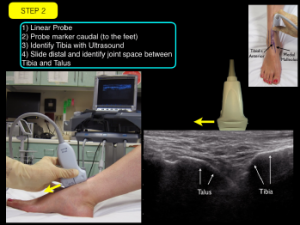
Stabilize the ultrasound transducer with your nondominant hand and have an 18–20-gauge 1.5 in. needle attached to a 5-cc syringe ready for aspiration. Because this is an out-of-plane technique, the operator will not be able visualize the needle entering the joint capsule. However, in our opinion, this is easier overall given the limited space of the tibiotalar joint. Puncture the skin just medial to the midline of the transducer using a very steep angle (just below 90º). The clinician should aspirate as the needle is inserted into the joint capsule because the needle tip may not be clearly visible (see Step 5).

Note: In the photographs for Steps 4 and 5, the sterile cover is not placed over the transducer. These images are to demonstrate transducer and needle positioning.


Summary

POCUS evaluation for joint capsule swelling of the ankle can be an important adjunct in the diagnostic evaluation of a patient with a painful and warm lower extremity. A simplified ultrasound technique for visualizing joint capsule swelling of the medial tibiotalar joint can be rapidly performed at bedside. With this critical information, the clinician can more confidently decide whether to perform an ultrasound-guided joint aspiration (or call a consultant for assistance) to determine the presence of a septic joint.


Dr. NagdevDr. Nagdev is director of emergency ultrasound at Highland General Hospital, Alameda Health System in Oakland, California.

References

  1. Movassaghi K, Wakefield C, Bohl DD, et al. Septic arthritis of the native ankle. JBJS Rev. 2019;7(3):e6.
  2. Roy S, Dewitz A, Paul I. Ultrasound-assisted ankle arthrocentesis. Am J Emerg Med. 1999;17(3):300-301.
  3. Wisniewski SJ, Smith J, Patterson DG, et al. Ultrasound-guided versus nonguided tibiotalar joint and sinus tarsi injections: A cadaveric study. PM R. 2010;2(4):277-281. 

Pages: 1 2 3 | Single Page

Topics: AnkleImaging & Ultrasoundjoint infectionsPOCUSPoint-of-Care Ultrasound

Related

  • ACEP’s October 2025 Poll: How Often Do You Read Your Own X-Rays?

    September 30, 2025 - 0 Comment
  • Case Report: Rapid Diagnosis of Acute Aortic Dissection with POCUS

    June 11, 2025 - 1 Comment
  • How Well Does Ultrasound Detect Volvulus in Infants?

    September 5, 2024 - 0 Comment

Current Issue

ACEP Now: November 2025

Download PDF

Read More

No Responses to “Using Point-of-Care Ultrasound to Evaluate and Aspirate Ankle Infections”

Leave a Reply Cancel Reply

Your email address will not be published. Required fields are marked *


*
*


Wiley
  • Home
  • About Us
  • Contact Us
  • Privacy
  • Terms of Use
  • Advertise
  • Cookie Preferences
Copyright © 2025 by John Wiley & Sons, Inc. All rights reserved, including rights for text and data mining and training of artificial technologies or similar technologies. ISSN 2333-2603