Logo

Log In Sign Up |  An official publication of: American College of Emergency Physicians
Navigation
  • Home
  • Multimedia
    • Podcasts
    • Videos
  • Clinical
    • Airway Managment
    • Case Reports
    • Critical Care
    • Guidelines
    • Imaging & Ultrasound
    • Pain & Palliative Care
    • Pediatrics
    • Resuscitation
    • Trauma & Injury
  • Resource Centers
    • mTBI Resource Center
  • Career
    • Practice Management
      • Benchmarking
      • Reimbursement & Coding
      • Care Team
      • Legal
      • Operations
      • Quality & Safety
    • Awards
    • Certification
    • Compensation
    • Early Career
    • Education
    • Leadership
    • Profiles
    • Retirement
    • Work-Life Balance
  • Columns
    • ACEP4U
    • Airway
    • Benchmarking
    • Brief19
    • By the Numbers
    • Coding Wizard
    • EM Cases
    • End of the Rainbow
    • Equity Equation
    • FACEPs in the Crowd
    • Forensic Facts
    • From the College
    • Images in EM
    • Kids Korner
    • Medicolegal Mind
    • Opinion
      • Break Room
      • New Spin
      • Pro-Con
    • Pearls From EM Literature
    • Policy Rx
    • Practice Changers
    • Problem Solvers
    • Residency Spotlight
    • Resident Voice
    • Skeptics’ Guide to Emergency Medicine
    • Sound Advice
    • Special OPs
    • Toxicology Q&A
    • WorldTravelERs
  • Resources
    • ACEP.org
    • ACEP Knowledge Quiz
    • Issue Archives
    • CME Now
    • Annual Scientific Assembly
      • ACEP14
      • ACEP15
      • ACEP16
      • ACEP17
      • ACEP18
      • ACEP19
    • Annals of Emergency Medicine
    • JACEP Open
    • Emergency Medicine Foundation
  • About
    • Our Mission
    • Medical Editor in Chief
    • Editorial Advisory Board
    • Awards
    • Authors
    • Article Submission
    • Contact Us
    • Advertise
    • Subscribe
    • Privacy Policy
    • Copyright Information

Using Point-of-Care Ultrasound to Evaluate and Aspirate Ankle Infections

By Arun Nagdev, MD | on January 21, 2020 | 0 Comment
Sound Advice
  • Tweet
  • Click to email a link to a friend (Opens in new window) Email
Print-Friendly Version

Septic arthritis of the ankle is an uncommon but devastating clinical entity representing 3 percent to 14 percent of all septic arthritis cases.1 Diagnostics can prove challenging, especially because systemic symptoms like fever and chills are present in fewer than 50 percent of cases. This forces clinicians to rely on nonspecific and often insensitive physical exam findings, such as pain with joint movement and axial loading.

You Might Also Like
  • Easy Ultrasound Technique to Evaluate and Aspirate an Atraumatic Painful Wrist
  • Ultrasound-Guided Elbow Arthrocentesis
  • Ultrasound-Guided Glenohumeral Joint Evaluation and Aspiration
Explore This Issue
ACEP Now: Vol 39 – No 01 – January 2020

In cases of clinical suspicion of a joint infection, point-of-care ultrasound (POCUS) is an ideal bedside tool to determine the presence of joint capsule effusion. Also, POCUS allows a safe and simplified method for joint aspiration.1–3

Ultrasound Examination of the Ankle

Using POCUS for ankle joint assessment starts with good positioning. Place the patient in the supine position with their foot in mild plantar flexion so that the sole rests against the bed. If possible, palpate the anterior tibialis tendon on the anteromedial aspect of the foot. This landmark is easily noted in most patients and acts as an easy starting point when using ultrasound to evaluate the tibiotalar joint.
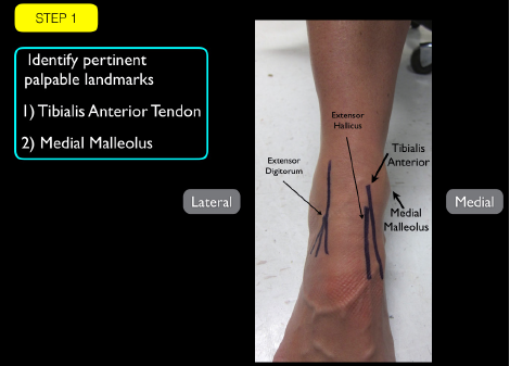
PHOTOS: Arun Nagdev

PHOTOS: Arun Nagdev

The goal is to image just under the anterior tibias tendon or just medial to it. The space between the anterior tibias tendon and medial malleolus is free from tendons or arteries, allowing for an unobstructed evaluation of the ankle joint as well as a safe location for aspiration. We recommend using a high-frequency linear transducer because of the shallow depth of the tibiotalar joint (see Step 1).

We also recommend imaging the nonaffected ankle first to determine a true normal. Place the transducer over the distal tibia (with the probe marker oriented caudally) and visualize the bright hyperechoic line of its bony cortex. Slowly slide the transducer toward the ankle until you are over the space between the tibia and talus (see Step 2). A joint capsule effusion will be an anechoic (dark) fluid collection just above the tibiotalar joint and is clearly seen on ultrasound. A superficial cellulitis and/or abscess can be easily differentiated from a deep joint effusion due to its location close to the probe and connection to the overlying skin (see Step 3).

In patients with an obvious effusion of the joint capsule overlying the tibiotalar joint, the clinician can progress to performing the ultrasound-guided aspiration or call a consultant for assistance. Clinical judgment is needed to determine the need for aspiration because other noninfectious entities can produce joint capsule swelling (eg, gout, arthritis, etc.).

Pages: 1 2 3 | Single Page

Topics: AnkleImaging & Ultrasoundjoint infectionsPOCUSPoint-of-Care Ultrasound

Related

  • ACEP’s October 2025 Poll: How Often Do You Read Your Own X-Rays?

    September 30, 2025 - 0 Comment
  • Case Report: Rapid Diagnosis of Acute Aortic Dissection with POCUS

    June 11, 2025 - 1 Comment
  • How Well Does Ultrasound Detect Volvulus in Infants?

    September 5, 2024 - 0 Comment

Current Issue

ACEP Now: November 2025

Download PDF

Read More

No Responses to “Using Point-of-Care Ultrasound to Evaluate and Aspirate Ankle Infections”

Leave a Reply Cancel Reply

Your email address will not be published. Required fields are marked *


*
*


Wiley
  • Home
  • About Us
  • Contact Us
  • Privacy
  • Terms of Use
  • Advertise
  • Cookie Preferences
Copyright © 2025 by John Wiley & Sons, Inc. All rights reserved, including rights for text and data mining and training of artificial technologies or similar technologies. ISSN 2333-2603