Figure 2: Schematic representation of the intercostal nerves as they travel from the thoracic spine. The distal lateral cutaneous branch exits at approximately the midaxillary line and pierces the internal intercostal muscle, external intercostal muscle, and serratus anterior muscle. The anterior fascial plane above the serratus anterior muscle acts as the target for this planar block.
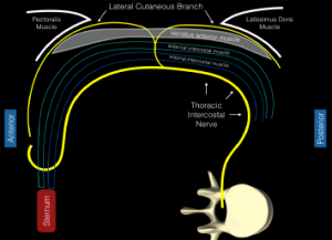
Figure 2: Schematic representation of the intercostal nerves as they travel from the thoracic spine. The distal lateral cutaneous branch exits at approximately the midaxillary line and pierces the internal intercostal muscle, external intercostal muscle, and serratus anterior muscle. The anterior fascial plane above the serratus anterior muscle acts as the target for this planar block.


No Responses to “Figure 2: Schematic representation of the intercostal nerves as they travel from the thoracic spine. The distal lateral cutaneous branch exits at approximately the midaxillary line and pierces the internal intercostal muscle, external intercostal muscle, and serratus anterior muscle. The anterior fascial plane above the serratus anterior muscle acts as the target for this planar block.”