2. Positioning
Explore This Issue
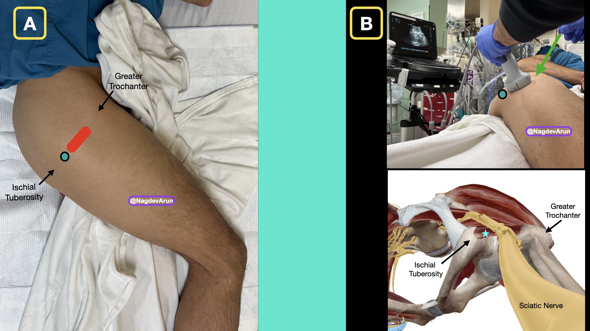
ACEP Now: Vol 41 – No 06 – June 2022PICTURE 2A (click to enlarge): Place the patient in a lateral position, if possible, with mild knee flexion. Note the probe (red oval) with the probe marker (green circle) pointed to the ischial tuberosity (medial). PICTURE 2B (click to enlarge): Representative anatomy when placing the ultrasound transducer in between the greater trochanter and ischial tuberosity. Note the inplane lateral to medial needle approach in the top image. Also, note the quadratus femoris muscle (blue star). Green circle indicates probe marker.
The patient’s proximal affected leg should be exposed. The patient should be in a lateral decubitus position with the affected side up and the knee flexed at a 90 degree angle. The ultrasound machine should be placed so that the clinician can look directly at the screen as well as the site of needle entry in the same line of sight (Pictures 2A and 2B).
3. Procedure Details
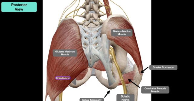
With the patient in the lateral decubitus position, palpate for the greater trochanter laterally and the posterior superior iliac spine medially. A line between the two landmarks will indicate the path of the needle. A curvilinear probe (1–5 MHz) should be placed along this line.
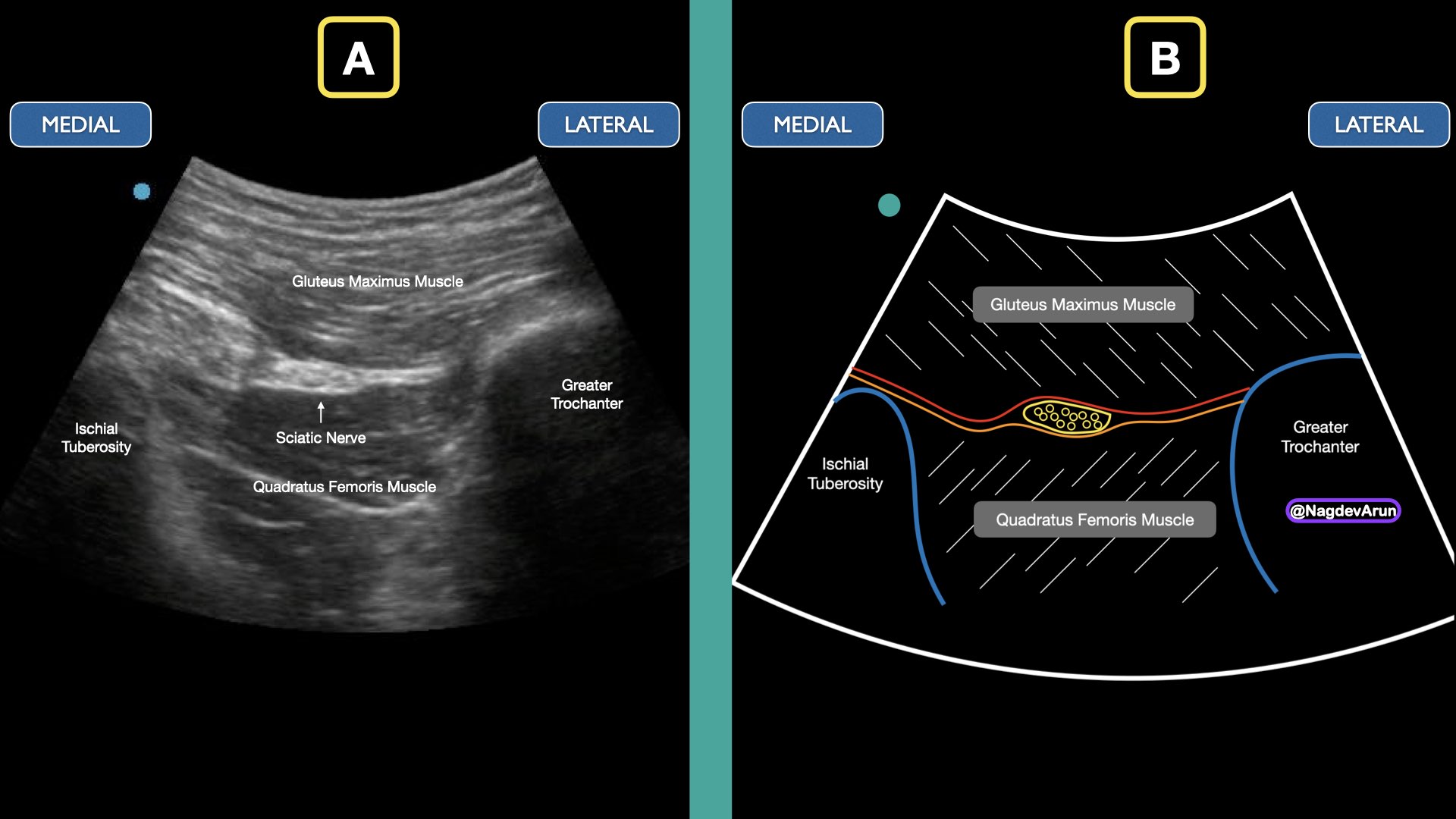
PICTURE 3A (click to enlarge): Ultrasound image for the TGSNB. Note the greater trochanter and ischial tuberosity. The sciatic nerve is located in the fascial planes between the gluteus maximus muscle (superficially) and the quadratus femoris muscle (deeper). PICTURE 3B (click to enlarge): Schematic representation of the ultrasound image.
The greater trochanter will be located laterally and the ischial tuberosity should be located medially identified as hyperechoic structures on the screen. In some patients, the ischial tuberosity will be hard to identify. The sciatic nerve will appear as a flattened, round or triangular hyperechoic structure lying within the fascial plane between the gluteus maximus and quadratus femoris muscles. It will usually be located about 4–6 cm below the skin. (Pictures 3A and 3B)
If having difficulty identifying the sciatic nerve at this level, you can also identify the distal sciatic nerve in the mid to distal posterior thigh and follow the nerve proximally to the level of the ischial tuberosity and greater trochanter. This video explains how to perform an ultrasound-guided distal sciatic nerve block in the popliteal fossa: https://www.acepnow.com/multimedia/ultrasound-guided-distal-sciatic-nerve-block/.
4. Skin Wheal
After satisfactory identification of the proximal sciatic nerve, widely prep the skin with chlorhexidine, allow to completely dry, and place 2–3 mL lidocaine skin wheal, 2–3 cm cephalad, or caudad to the transducer. The site can be either cephalad or caudad to the probe as the sciatic nerve can be approached from either direction. After sterile prep, place a lidocaine skin wheal at the insertion site using a 27 gauge needle.
Pages: 1 2 3 4 | Single Page


No Responses to “How To Relieve Sciatica Pain with New TransGluteal Nerve Block Treatment”