Evaluation
When evaluating a Weber B fracture, if the initial imaging does not demonstrate obvious evidence of tibiotalar instability, ankle stress testing is indicated. There are three primary methods of performing an ankle stress test. These include manual, gravity, and weight-bearing techniques.
Explore This Issue
ACEP Now: Vol 39 – No 04 – April 2020Manual stress testing has historically been the method utilized to evaluate the stability of the ankle joint. This method involves keeping the ankle at neutral dorsiflexion, rotating the tibia internally at 10°, and applying 8 to 20 pounds of external rotation at the foot.3 This typically requires a physician going to the radiology suite to perform the stress test.
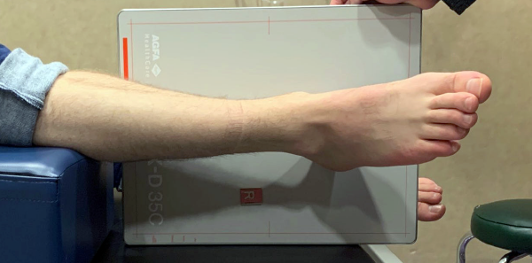
Figure 1: Optimal ankle positioning with gravity stress testing. Joseph Noack & Spencer Tomberg.
Gravity stress is typically performed with the patient lying in lateral decubitus with the injured side down, allowing the foot and ankle to create a lateral force across the ankle joint, with the foot resting in natural plantar flexion and the leg internally rotated at 10° to 15° (see Figure 1).6 Gravity stress has been shown to be as reliable and less painful than manual stress testing.6,7
Weight-bearing films are a relatively new method of testing for medial stability. Despite initial cadaveric studies demonstrating that weight-bearing films did not accurately provide radiographic evidence of instability, recent clinical studies have shown that weight-bearing radiographs are predictive of stability and that gravity stress radiographs likely overestimate the instability, resulting in up to a tenfold increase in surgeries when a medial clear space (MCS) cutoff of 4 mm is used.8–11 One recent study compared patients who had a borderline unstable ankle based on gravity stress imaging (MCS 4–7 mm) but stability on the weight-bearing imaging to patients who had a stable ankle based on gravity and weight-bearing imaging. There was no functional outcome difference between the two cohorts of patients who elected to be managed nonoperatively.10 The most compelling argument for weight-bearing films is that they stress the ankle joint under physiological conditions that measure stability under realistic and reproducible conditions.
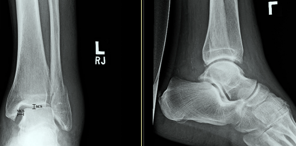
Figure 2: Normal ankle mortise view with demonstrated medial clear space (MCS) and superior clear space (SCS).
Joseph Noack & Spencer Tomberg
Like gravity stress imaging, weight-bearing films can be performed by a radiology technician without physician assistance. The weight-bearing technique is more reproducible and takes less radiology technician training than either gravity stress or manual stress views.10 However, from an emergency medicine perspective, one of the most glaring questions is whether patients with an acutely injured ankle can bear enough weight to get adequate radiographs, as the studies comparing stress techniques have been completed in orthopedic clinics three to 10 days after the initial injury. We could not find data pertaining to this particular question.
MRI can be used to evaluate the deltoid ligament, but the degree of the tear does not always equate to instability on stress radiographs.12
Specific parameters are evaluated in each stress view of the ankle. For gravity stress views, consensus is leaning to a value for the MCS of <7 mm to define a stable ankle joint. An MCS >4 mm is the historical value used to indicate operative management, but this value has been shown to lead to a high false-positive rate and unnecessary surgeries, of which 10 percent have surgical complications.13,14 An MCS greater than the superior clear space (SCS) of 1 mm or more on mortise view is another sign of ankle instability (see Figure 2). Finally, if the injured side has an MCS that is >2 mm wider than the uninjured side, the ankle can be considered unstable. Any of these radiographic abnormalities on initial imaging suggest a clinically significant injury to the deltoid ligament and ankle instability.13 While the trend is toward adopting the 6 mm threshold for MCS, there is no consensus among orthopedic surgeons and your consultant may use stricter guidelines.6,13
Pages: 1 2 3 4 | Single Page


One Response to “Tips for Managing Weber B Ankle Fractures”
May 19, 2020
Huda NaeemIs there an app for this ACEPNOW?