Logo

Log In Sign Up |  An official publication of: American College of Emergency Physicians
Navigation
  • Home
  • Multimedia
    • Podcasts
    • Videos
  • Clinical
    • Airway Managment
    • Case Reports
    • Critical Care
    • Guidelines
    • Imaging & Ultrasound
    • Pain & Palliative Care
    • Pediatrics
    • Resuscitation
    • Trauma & Injury
  • Resource Centers
    • mTBI Resource Center
  • Career
    • Practice Management
      • Benchmarking
      • Reimbursement & Coding
      • Care Team
      • Legal
      • Operations
      • Quality & Safety
    • Awards
    • Certification
    • Compensation
    • Early Career
    • Education
    • Leadership
    • Profiles
    • Retirement
    • Work-Life Balance
  • Columns
    • ACEP4U
    • Airway
    • Benchmarking
    • Brief19
    • By the Numbers
    • Coding Wizard
    • EM Cases
    • End of the Rainbow
    • Equity Equation
    • FACEPs in the Crowd
    • Forensic Facts
    • From the College
    • Images in EM
    • Kids Korner
    • Medicolegal Mind
    • Opinion
      • Break Room
      • New Spin
      • Pro-Con
    • Pearls From EM Literature
    • Policy Rx
    • Practice Changers
    • Problem Solvers
    • Residency Spotlight
    • Resident Voice
    • Skeptics’ Guide to Emergency Medicine
    • Sound Advice
    • Special OPs
    • Toxicology Q&A
    • WorldTravelERs
  • Resources
    • ACEP.org
    • ACEP Knowledge Quiz
    • Issue Archives
    • CME Now
    • Annual Scientific Assembly
      • ACEP14
      • ACEP15
      • ACEP16
      • ACEP17
      • ACEP18
      • ACEP19
    • Annals of Emergency Medicine
    • JACEP Open
    • Emergency Medicine Foundation
  • About
    • Our Mission
    • Medical Editor in Chief
    • Editorial Advisory Board
    • Awards
    • Authors
    • Article Submission
    • Contact Us
    • Advertise
    • Subscribe
    • Privacy Policy
    • Copyright Information

Tips for Diagnosing Occult Fractures in the Emergency Department

By Arun Sayal, MD, CCFP(EM) | on September 19, 2018 | 0 Comment
Casted
  • Tweet
  • Click to email a link to a friend (Opens in new window) Email
Print-Friendly Version

Imagine that on your next shift, you see two patients with hip pain and normal X-rays. Patient A is a 25-year-old male who fell down two stairs onto his right hip. He has a limp and lateral hip tenderness, but he has a good range of motion (neither shortened or externally rotated). His estimated pretest probability for a fracture is 10 percent.

You Might Also Like
  • Tips for Emergency Physicians on Spotting Occult Knee Dislocation
  • Emergency Medicine Pearls, Pitfalls for Treatment of Pediatric Distal Radius Fractures
  • When to Use Point-of-Care Ultrasound for Skull Fractures
Explore This Issue
ACEP Now: Vol 37 – No 09 – September 2018

Patient B is a 78-year-old male who fell on his right hip from standing. He can’t walk due to the pain, is tender to palpation laterally at the hip, and has quite reduced active and passive range of motion of the hip due to pain (neither shortened or externally rotated). His estimated pretest probability for a fracture is 90 percent.

Again, both have normal X-rays of the hip and pelvis.

You mark Patient A’s 10 percent pretest probability of a fracture on a Fagan nomogram on the left axis, then draw a line from that mark through the estimated LR of 0.075 on the center axis and see where it intersects on post-test probability (ie, the right axis). See the blue line on Figure 1. Given the negative X-ray, you see the post-test probability of fracture is now less than 1 percent.

You do the same for Patient B, and despite his pretest probability of 90 percent for a fracture, you see the negative X-ray has driven the post-test probability of fracture down to around 40 percent. See the red line on Figure 1.

Negative X-rays in the face of a low pretest probability are unlikely to have a fracture, of course. However, we should keep the possibility of a fracture higher on the differential diagnosis for patients with a higher pretest probability for fracture and negative X-rays.

Remember this axiom: The purpose of any test is not to make the diagnosis but rather to affect our pre-test probability.

Case Resolutions

Case 1: occult mid-shaft clavicle fracture

The 12-year-old boy who fell on his shoulder: When he returned one week later he was still tender and swollen and looked like he had a clavicle fracture. He was kept in a sling, and X-rays repeated three weeks later revealed the fracture.

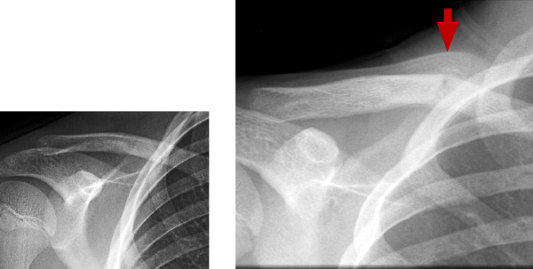
Left: Day 1 – normal
Right: Week 3 – fracture and callus

Case 2: occult distal tibia fracture

The 13-year-old girl with a twisted ankle: When she returned one week later, her mechanism was external rotation (a red flag, see image), and she was tender over her distal tibia. X-rays taken three weeks later showed an occult distal tibia fracture.

Pages: 1 2 3 4 | Single Page

Topics: BonesfractureImaging & UltrasoundX-Ray

Related

  • Patterns of Injury in Elder Abuse

    November 6, 2025 - 0 Comment
  • Poll: Emergency Physicians Read Their Own X-Rays More Often Than Not

    November 6, 2025 - 0 Comment
  • ACEP’s October 2025 Poll: How Often Do You Read Your Own X-Rays?

    September 30, 2025 - 0 Comment

Current Issue

ACEP Now: November 2025

Download PDF

Read More

No Responses to “Tips for Diagnosing Occult Fractures in the Emergency Department”

Leave a Reply Cancel Reply

Your email address will not be published. Required fields are marked *


*
*


Wiley
  • Home
  • About Us
  • Contact Us
  • Privacy
  • Terms of Use
  • Advertise
  • Cookie Preferences
Copyright © 2025 by John Wiley & Sons, Inc. All rights reserved, including rights for text and data mining and training of artificial technologies or similar technologies. ISSN 2333-2603