Explore This Issue
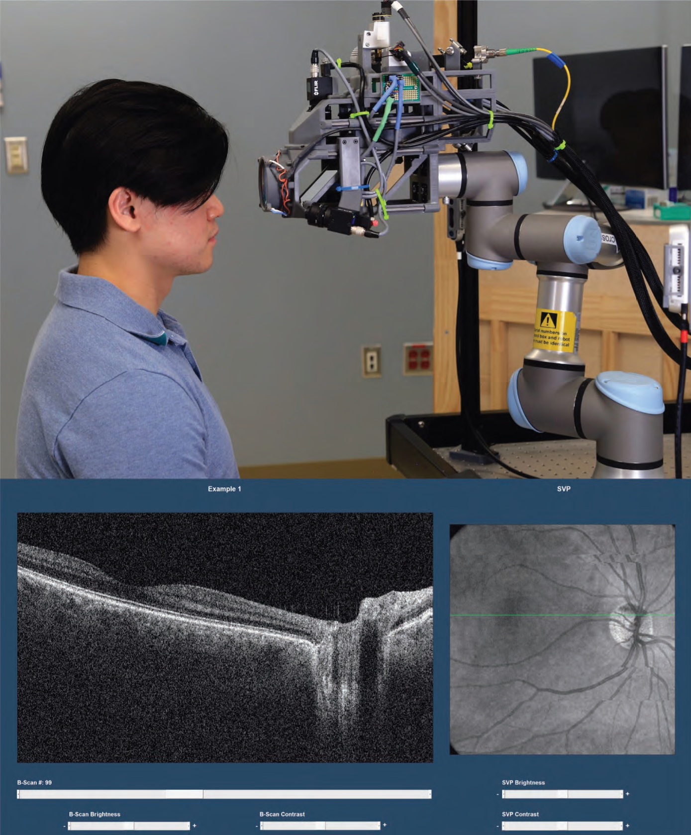
ACEP Now: Vol 42 – No 03 – March 2023FIGURE 1: Robotic optical coherence tomography (OCT) device (top) and screenshot of OCT viewing software (bottom) showing a cross-sectional OCT image (left) at the level of the green line in the head-on projection (right), which shows the view of fundus examination. (Click to enlarge.)
Importantly, the study suggests that OCT may be of real diagnostic value to emergency physicians. Though most emergency physicians are likely not familiar with the interpretation of OCT images, the nine emergency physicians who participated in the study quickly learned to differentiate abnormal from normal images after a 30-minute training session on the basics of ocular OCT. They were able to interpret the OCT images with 100 percent sensitivity for urgent or emergent abnormalities and 69 percent sensitivity for any posterior eye abnormality when evaluated against reference-standard diagnosis based on a combination of ophthalmology-consultation diagnosis and retina-specialist OCT review. These results were in stark contrast to emergency physician-performed direct ophthalmoscopy, which did not detect any abnormalities in the same patients. As patients with urgent and emergent eye conditions often experience delays in care and subsequent worse outcomes in current clinical practice, these pilot study results indicate that this new technology has the potential to substantially improve the quality of acute eye care by facilitating more accurate ophthalmology referral decisions.
Among the 72 eyes imaged with the robotic OCT device, emergency physicians assessed a broad range of urgent and emergent retinal and optic nerve pathologies such as papilledema, retinal detachment, and retinal vascular occlusion. The three-dimensional nature of OCT imaging makes the detection of these pathologies substantially easier, compared with the two-dimensional view provided by fundus examination. For example, optic disc edema that looks subtle on fundus examination can be obvious on OCT, as elevation of the optic nerve head caused by swelling is readily appreciable in cross sections.3
Currently, the robotic OCT device is still investigational and not commercially available, but this novel technology shows promise in expanding the ability of emergency physicians to diagnose conditions affecting the posterior eye accurately and efficiently.
 Dr. Song is a fourth-year medical student at Duke University. Her research focuses on developing and translating innovative technologies such as robotics and machine learning to improve eye care in non-ophthalmology settings including the emergency department.
 Dr. Kuo is an associate professor of ophthalmology and assistant professor of biomedical engineering at Duke University.
References
- Mackay DD, Garza PS, Bruce BB, Newman NJ, Biousse V. The demise of direct ophthalmoscopy: A modern clinical challenge. Neurol Clin Pract. 2015;5(2):150-157.
- Song A, Roh KM, Lusk JB, et al. Robotic optical coherence tomography retinal imaging for emergency department patients: a pilot study for emergency physicians’ diagnostic performance. Ann Emerg Med. 2023;S0196-0644(22)01196-9.
- Wang JK, Kardon RH, Kupersmith MJ, Garvin MK. Automated quantification of volumetric optic disc swelling in papilledema using spectral-domain optical coherence tomography. Investigative Ophthalmology & Visual Science. 2012;53(7):4069-4075.
Pages: 1 2 3 | Single Page


No Responses to “The Real-World Utility of Ophthalmic Tomography”