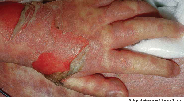
All skin and mucous membranes are susceptible to involvement. Mucous membrane involvement often precedes skin lesions, appearing with the prodromal illness (eg, fever, cough, sore throat). The rash often begins as a poorly defined erythematous macular rash that progresses in hours to days. This patient has TEN “without spots,” which is the less-common form of TEN, lacking target lesions. This is compared to TEN “with spots,” which features widespread erythematous or purpuric macules with blistering on the macule.2
Explore This Issue
ACEP Now: Vol 34 – No 02 – February 2015Drugs are most often implicated in TEN. One study at a major pediatric burn intensive care unit cited medication use in all 15 described cases.3 The most common drugs cited are antibacterials, sulfonamides, nonsteroidal anti-inflammatory drugs, allopurinol, disease-modifying agents, anti-epileptics, anti-retroviral agents, and corticosteroids.4 Infectious causes including mycoplasma and herpes virus have also been documented.
Are there any confirmatory tests for TEN that can be performed in the emergency department?
- The diagnosis of TEN is generally clinical. The patient’s history (particularly medication history) and physical exam (particularly head, eyes, ears, nose, throat, genitourinary, and skin) are imperative.
- The only confirmatory test is histopathologic analysis of a skin biopsy; full thickness epithelial necrosis is diagnostic.
- There is no definitive laboratory or imaging study to make the diagnosis.
- As the disease progresses with multiorgan involvement, more laboratory test abnormalities may be identified.
What clinical clues suggest the diagnosis of TEN in a child?
- Increased salivation and decreased appetite from oral involvement.
- Painful micturition due to urethral involvement.
- Profuse diarrhea due to enteral involvement.
- Respiratory distress due to involvement of the bronchial epithelium.
- Ocular lesions, which may indicate a higher chance for morbidity due to tearing of the epidermis leading to conjunctival erosions, keratitis, and corneal erosions.
What else should be considered when treating patients diagnosed with TEN?
Differential diagnosis should include Kawasaki disease, staphylococcal scalded skin syndrome, toxic shock syndrome, disseminated adenovirus infection, infectious conjunctivitis, Reiter’s syndrome, hypersensitivity vasculitis, erythema multiforme, exfoliative conjunctivitis, disseminated herpes infection, and measles.
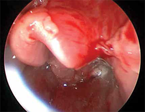
Figure 1: Supraglottic desquamation of lingual surface of epiglottis and vallecula.

Figure 2: The patient’s trachea appears normal.
What immediate threats must be addressed in a patient with TEN?
In the ED, the treatment of the patient with suspected SJS/TEN is akin to that of a patient with severe burns.
- Discontinue any identified offending agent early.
- Consider early transfer to a burn unit because these interventions are associated with improved outcomes.5
- Early fluid resuscitation is imperative in any patient who appears to be in shock.
- Broad-spectrum antibiotic administration is warranted if the child meets criteria for sepsis or if infectious etiology of symptoms is considered likely; however, routine antibiotic administration is not indicated as in the case of severe burns.
- Manage fluids and electrolytes, provide adequate analgesia, prevent secondary infection, and keep the patient warm.
Other than supportive care, what can be done for a patient with TEN?6
Therapeutic measures for TEN remain controversial.
- Most dermatologists at this time do not advocate corticosteroid use.
- Intravenous immunoglobulin (IVIG) is often utilized in treatment protocols, but no randomized controlled trials to date have shown definitive benefit.
Case Resolution
While the patient is on the floor, an echocardiogram is scheduled for the next day to check for potential Kawasaki disease. Infectious disease and dermatology are consulted and scheduled to see the patient the next day. Urology is consulted for placement of a urethral catheter due to lack of urine output and urethral pain. By the eighth hour of hospital admission, the rash has quickly changed quality from macular to bullous and vesicular and has spread to involve the majority of surfaces of his body, including the palms and soles. The patient is transferred to the pediatric ICU with a new oxygen requirement of 0.5 L by nasal cannula and concern for upper airway involvement with TEN.
Pages: 1 2 3 4 | Single Page


No Responses to “Recognize Pediatric Toxic Epidermal Necrolysis Symptoms, Manage Disease”