Anesthetic: For the anesthetic, we recommend lidocaine 1 to 2 percent, bupivacaine 0.25 percent, 2 to 5 cc per nerve block. For the less experienced provider, we recommend starting with lidocaine, as bupivacaine is more commonly associated with local anesthetic systemic toxicity. All anesthetics should be used with caution when injecting near vascular structures, with LipidRescue available if needed.
Explore This Issue
ACEP Now: Vol 35 – No 10 – October 2016Positioning: Position the ultrasound system across from the affected extremity at a comfortable height. Place the patient in a supine or 45-degree upright position, with the affected arm supinated and externally rotated and the forearm resting on a stand. As you start with the linear ultrasound probe in a transverse position in the distal forearm, align the probe marker to the same side as it appears to you on your screen. The provider should have both the ultrasound screen and the point of needle entry in the same line of sight if possible.
Locating Each Nerve
The median nerve lies along the midline of the forearm in the fascial plane between the flexor digitorum superficialis and profundus tendons. Place the linear ultrasound probe in a transverse position at the wrist crease on the volar surface of the distal forearm. Scan proximally to the mid-forearm and look for the classic “honeycomb” appearance of a nervous structure at the junction of several fascial planes (see Figure 3). There are no associated vascular structures, except in rare incidences. In the distal wrist, muscle tendon sheaths will look similar to the classic honeycomb appearance of the nerves. By scanning proximally, tendons will disappear and the nerve will be clearer.
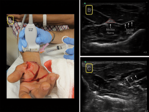
Figure 3. For the median nerve block, place the linear ultrasound probe in a transverse position at the wrist crease on the volar surface of the distal forearm. Scan proximally to the mid-forearm (A) and look for the classic “honeycomb” appearance of a nervous structure at the junction of several fascial planes (B). insert your needle from whichever side is most comfortable (A&C).

Figure 4. For the radial nerve block, starting at the wrist with the linear ultrasound probe in a transverse position over the radial artery (A), scan proximally until the nerve can be visualized separating from the radial artery (B). Locate the radial nerve between the brachioradialis and brachialis muscles. insert your needle from the radial (lateral) aspect of the probe (A&C).
The radial nerve can be found in a position radial (lateral) to the radial artery. Starting at the wrist with the linear ultrasound probe in a transverse position over the radial artery, scan proximally until the nerve can be visualized separating from the radial artery (see Figure 4). Alternatively, you can slide the probe proximally and find the nerve in the supracondylar area. Keeping the probe transverse on the lateral aspect of the arm, locate the radial nerve between the brachioradialis and brachialis muscles. You can perform the block from this position or trace the nerve back distally to a comfortable position in the forearm.
Pages: 1 2 3 4 | Single Page


No Responses to “How to Perform Ultrasound-Guided Forearm Nerve Blocks to Provide Non-Drug Pain Relief for Acute Injuries”