Identify and Treat Lisfranc Injuries
Explore This Issue
ACEP Now: Vol 36 – No 12 – December 2017Figure 2: The Lisfranc ligament connects the base of the second metatarsal to the lateral aspect of the medial cuneiform.
Credit: Siddiqui N A, Galizia M S, Almusa E, et al. RadioGraphics. 2014;34(2):514-531.
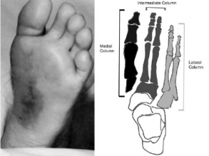
Figure 3: Left: Plantar ecchymosis. Right: Medial, intermediate and lateral column associated with midfoot biomechanics.
Emerg Med J. 2017;34:52-56.
Physical Exam Suggestive of Lisfranc Fracture
- Inability to bear weight or stand on tiptoes.2
- Ecchymosis on medial plantar aspect of foot is pathognomonic but may be absent in minor fractures (see Figure 3).2
- Dorsal midfoot pain and swelling.
- Pain elicited with passive supination and pronation of the forefoot with the hindfoot held fixed.4
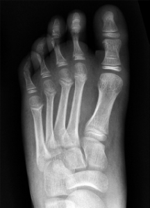
Figure 4A: Initial AP non-weight-bearing radiograph shows normal alignment of the medial and middle columns of an 11-year-old boy after a fall.
Credit: Siddiqui N A, Galizia M S, Almusa E, et al. RadioGraphics. 2014;34(2):514-531.
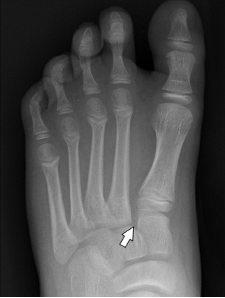
Figure 4b: AP weight-bearing radiograph shows widening of the joint seen as a gap of more than 2 mm between C1 and M2 and between M1 and M2 (arrow).
Credit: Siddiqui N A, Galizia M S, Almusa E, et al. RadioGraphics. 2014;34(2):514-531.
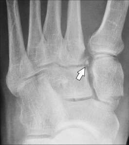
Figure 6: AP weight-bearing radiograph showing a small chip fracture from medial margin of base of M2, known as the “fleck sign.”
Credit: Siddiqui N A, Galizia M S, Almusa E, et al. RadioGraphics. 2014;34(2):514-531.
Diagnosis
Obtain three view radiographs of the foot (anteroposterior [AP], lateral, and standard 45-degree oblique views). Ideally, weight-bearing stress views should be obtained since initial plain X-rays may fail to show subtle widening of the articulation spaces (See Figure 4).1 Consider a CT scan of the foot if X-rays do not show an injury but you remain highly suspicious.
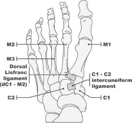
Normal Three-Column Anatomy of Lisfranc Complex on X-Ray (see Figures 1 and 5)
- On the AP view, the medial edge of the base of the second metatarsal (M2) should line up with the medial edge of the middle cuneiform (C2).1
- The gap between the second metatarsal and medial cuneiform is <2 mm.
- On the oblique view, the medial edge of the third and fourth metatarsal should line up with the medial edges of the middle cuneiform and cuboid, respectively.1
- On the lateral view, the superior border of the first metatarsal (M1) should align with the superior border of the medial cuneiform (C1).
Pages: 1 2 3 | Single Page


No Responses to “Identify and Treat Lisfranc Injuries”