Logo

Log In Sign Up |  An official publication of: American College of Emergency Physicians
Navigation
  • Home
  • Multimedia
    • Podcasts
    • Videos
  • Clinical
    • Airway Managment
    • Case Reports
    • Critical Care
    • Guidelines
    • Imaging & Ultrasound
    • Pain & Palliative Care
    • Pediatrics
    • Resuscitation
    • Trauma & Injury
  • Resource Centers
    • mTBI Resource Center
  • Career
    • Practice Management
      • Benchmarking
      • Reimbursement & Coding
      • Care Team
      • Legal
      • Operations
      • Quality & Safety
    • Awards
    • Certification
    • Compensation
    • Early Career
    • Education
    • Leadership
    • Profiles
    • Retirement
    • Work-Life Balance
  • Columns
    • ACEP4U
    • Airway
    • Benchmarking
    • Brief19
    • By the Numbers
    • Coding Wizard
    • EM Cases
    • End of the Rainbow
    • Equity Equation
    • FACEPs in the Crowd
    • Forensic Facts
    • From the College
    • Images in EM
    • Kids Korner
    • Medicolegal Mind
    • Opinion
      • Break Room
      • New Spin
      • Pro-Con
    • Pearls From EM Literature
    • Policy Rx
    • Practice Changers
    • Problem Solvers
    • Residency Spotlight
    • Resident Voice
    • Skeptics’ Guide to Emergency Medicine
    • Sound Advice
    • Special OPs
    • Toxicology Q&A
    • WorldTravelERs
  • Resources
    • ACEP.org
    • ACEP Knowledge Quiz
    • Issue Archives
    • CME Now
    • Annual Scientific Assembly
      • ACEP14
      • ACEP15
      • ACEP16
      • ACEP17
      • ACEP18
      • ACEP19
    • Annals of Emergency Medicine
    • JACEP Open
    • Emergency Medicine Foundation
  • About
    • Our Mission
    • Medical Editor in Chief
    • Editorial Advisory Board
    • Awards
    • Authors
    • Article Submission
    • Contact Us
    • Advertise
    • Subscribe
    • Privacy Policy
    • Copyright Information

How To: The Ultrasound-Guided Supraclavicular Brachial Plexus Block

By Alex Ayala, MD; Henry Ashworth, MD, MPH; Arun Nagdev, MD | on April 6, 2023 | 0 Comment
Sound Advice
  • Tweet
  • Click to email a link to a friend (Opens in new window) Email
Print-Friendly Version

The clinician should clearly visualize the fluid deposition on the ultrasound screen, and use this fluid to lift the nerve bundle away from the first rib (Figure 4). The clinician should then deposit small aliquots (3 to 5 cc max) every 15 to 30 seconds until the predetermined total anesthetic is deposited. If the needle location is adequate, a single injection should suffice, but repositioning the needle superiorly may be necessary to surround the nerve sheath completely with anesthetic. Once your injection is complete, gently remove the needle and dress the injection site as needed.

You Might Also Like
  • Ultrasound-Guided Interscalene Approach To the Brachial Plexus Nerve Block
  • Pain Control Using Ultrasound-Guided Superficial Cervical Plexus Block
  • How to Perform Ultrasound-Guided Interscalene Nerve Blocks
Explore This Issue
ACEP Now: Vol 42 – No 04 – April 2023

Aftercare

Mark the affected extremity with the date, time, and type of block performed. Document the block in the electronic health record. Communicate with nurse(s), other providers, and any relevant consultants that a block was performed. Reassess the patient to ensure adequate analgesia and assess the efficacy of the block. Also, reassess and document a repeat neurovascular examination.

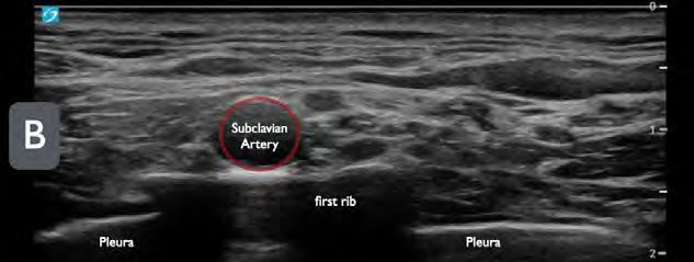
FIGURE 4A: Schematic representation of a block needle entering lateral to medial in-plane and depositing anesthetic between the brachial plexus and first rib. (Click to enlarge.)

Conclusion

For upper extremity injuries, traditionally procedural sedation, local anesthesia, and hematoma blocks have been used for pain management during procedures in the ED. However, these strategies have their own disadvantages such as increased resource allocation for procedural sedation and possibly incomplete analgesia for hematoma blocks depending on the location. Brachial plexus blocks such as the supraclavicular block provide adequate analgesia for a larger variety of upper arm injuries when compared to the hematoma block and require fewer resources when compared to procedural sedation. This nerve block is safe, easily learned, and does not require advanced ultrasound training to perform.11

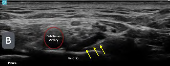
FIGURE 4B: Ultrasound image of a supraclavicular brachial plexus block. (yellow arrows = block needle). Note the anechoic (black) fluid between the first rib and the brachial plexus. (Click to enlarge.)

With an increase in the number of patients boarding in the ED, potentially decreased availability of procedural sedation rooms, and increasing patient-to-nurse ratios, ED physicians must adapt and become more efficient with limited resources. The supraclavicular block will not solve these systemic issues, but rather provides a more efficient strategy to manage pain better from acute upper extremity injuries as well as an alternative to procedural sedation in the ED.


Dr. Ayala is an emergency medicine resident at Highland General Hospital, Alameda Health System in Oakland, California.

Pages: 1 2 3 4 5 | Single Page

Topics: ClinicalImaging & UltrasoundNerve Blocks

Related

  • Non-Invasive Positive Pressure Ventilation in the Emergency Department

    October 1, 2025 - 0 Comment
  • Emergency Department Management of Prehospital Tourniquets

    October 1, 2025 - 0 Comment
  • ACEP’s October 2025 Poll: How Often Do You Read Your Own X-Rays?

    September 30, 2025 - 0 Comment

Current Issue

ACEP Now: November 2025

Download PDF

Read More

No Responses to “How To: The Ultrasound-Guided Supraclavicular Brachial Plexus Block”

Leave a Reply Cancel Reply

Your email address will not be published. Required fields are marked *


*
*


Wiley
  • Home
  • About Us
  • Contact Us
  • Privacy
  • Terms of Use
  • Advertise
  • Cookie Preferences
Copyright © 2025 by John Wiley & Sons, Inc. All rights reserved, including rights for text and data mining and training of artificial technologies or similar technologies. ISSN 2333-2603