Logo

Log In Sign Up |  An official publication of: American College of Emergency Physicians
Navigation
  • Home
  • Multimedia
    • Podcasts
    • Videos
  • Clinical
    • Airway Managment
    • Case Reports
    • Critical Care
    • Guidelines
    • Imaging & Ultrasound
    • Pain & Palliative Care
    • Pediatrics
    • Resuscitation
    • Trauma & Injury
  • Resource Centers
    • mTBI Resource Center
  • Career
    • Practice Management
      • Benchmarking
      • Reimbursement & Coding
      • Care Team
      • Legal
      • Operations
      • Quality & Safety
    • Awards
    • Certification
    • Compensation
    • Early Career
    • Education
    • Leadership
    • Profiles
    • Retirement
    • Work-Life Balance
  • Columns
    • ACEP4U
    • Airway
    • Benchmarking
    • Brief19
    • By the Numbers
    • Coding Wizard
    • EM Cases
    • End of the Rainbow
    • Equity Equation
    • FACEPs in the Crowd
    • Forensic Facts
    • From the College
    • Images in EM
    • Kids Korner
    • Medicolegal Mind
    • Opinion
      • Break Room
      • New Spin
      • Pro-Con
    • Pearls From EM Literature
    • Policy Rx
    • Practice Changers
    • Problem Solvers
    • Residency Spotlight
    • Resident Voice
    • Skeptics’ Guide to Emergency Medicine
    • Sound Advice
    • Special OPs
    • Toxicology Q&A
    • WorldTravelERs
  • Resources
    • ACEP.org
    • ACEP Knowledge Quiz
    • Issue Archives
    • CME Now
    • Annual Scientific Assembly
      • ACEP14
      • ACEP15
      • ACEP16
      • ACEP17
      • ACEP18
      • ACEP19
    • Annals of Emergency Medicine
    • JACEP Open
    • Emergency Medicine Foundation
  • About
    • Our Mission
    • Medical Editor in Chief
    • Editorial Advisory Board
    • Awards
    • Authors
    • Article Submission
    • Contact Us
    • Advertise
    • Subscribe
    • Privacy Policy
    • Copyright Information

How To: The Ultrasound-Guided Supraclavicular Brachial Plexus Block

By Alex Ayala, MD; Henry Ashworth, MD, MPH; Arun Nagdev, MD | on April 6, 2023 | 0 Comment
Sound Advice
  • Tweet
  • Click to email a link to a friend (Opens in new window) Email
Print-Friendly Version

Sonoanatomy

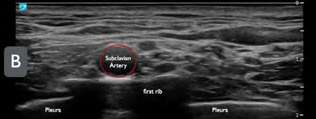
Place the high-frequency linear transducer above the upper border of the mid-to-lateral clavicle in a coronal oblique plane (parallel to the clavicle and aiming the beam into the thorax) (Figure 1B). Move the transducer medially to find the anechoic, round, and pulsatile subclavian artery (can be confirmed by color Doppler). The brachial plexus often appears superior and lateral to the artery, and often looks like a “cluster of grapes.” Note the first rib, a hyperechoic line with distal shadowing just under the brachial plexus. This should be contrasted to the normal lung sliding in the far field (Figure 2). In many patients, the first rib may not be clearly seen and the clinician will have to rotate the tail of the transducer in a counterclockwise manner to bring this sonographic landmark into view (Figure 3).

You Might Also Like
  • Ultrasound-Guided Interscalene Approach To the Brachial Plexus Nerve Block
  • Pain Control Using Ultrasound-Guided Superficial Cervical Plexus Block
  • How to Perform Ultrasound-Guided Interscalene Nerve Blocks
Explore This Issue
ACEP Now: Vol 42 – No 04 – April 2023

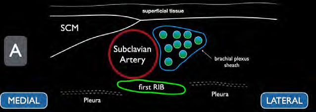
FIGURE 2A: Schematic representation of the supraclavicular brachial plexus. (Click to enlarge.)

An alternative technique, should you have difficulty finding the brachial plexus, is to start in the neck by identifying the carotid artery and internal jugular vein about the level of the cricoid cartilage.8 Slide the probe inferiorly toward the clavicle, reducing the angle between the probe and the skin as the probe is moved. In addition to using the subclavian vessels as a landmark, also be sure to identify any other vascular structures in the area of interest. After locating the brachial plexus and the projected needle path, we recommend use of color Doppler to prevent inadvertent vascular puncture.10

FIGURE 2B: Ultrasound image of the supraclavicular brachial plexus. (Click to enlarge)

Performing the Block

Once the optimal transducer position and needle path is identified, place a small skin wheal of local anesthetic just lateral to the transducer to anesthetize the overlying skin. After properly disinfecting the skin, enter through the skin wheal for a lateral to medial in-plane approach with the nerve block needle. Ensure continuous needle visualization and advance the needle tip until it touches the first rib. The goal is to find the area between the first rib and nerve bundle to inject anesthetic. Once you have clear needle-tip visualization within this space and have gently aspirated to ensure the needle tip is not intravascular, we recommend gently injecting a small amount of normal saline to ensure proper spread of anechoic fluid (hydrodissection).

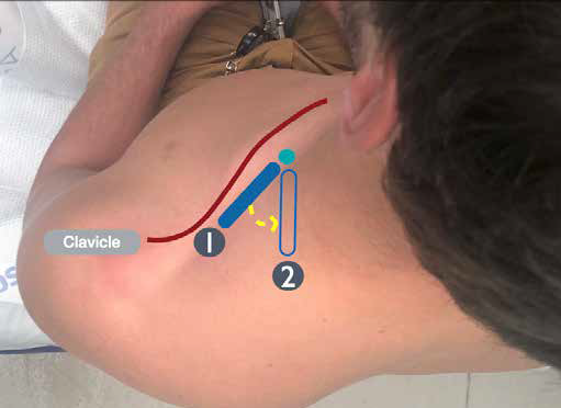
FIGURE 3: By rotating the ultrasound probe in a counterclockwise manner, the first rib can often be seen more clearly in cross-section on ultrasound. (Click to enlarge.)

Pages: 1 2 3 4 5 | Single Page

Topics: ClinicalImaging & UltrasoundNerve Blocks

Related

  • Non-Invasive Positive Pressure Ventilation in the Emergency Department

    October 1, 2025 - 0 Comment
  • Emergency Department Management of Prehospital Tourniquets

    October 1, 2025 - 0 Comment
  • ACEP’s October 2025 Poll: How Often Do You Read Your Own X-Rays?

    September 30, 2025 - 0 Comment

Current Issue

ACEP Now: November 2025

Download PDF

Read More

No Responses to “How To: The Ultrasound-Guided Supraclavicular Brachial Plexus Block”

Leave a Reply Cancel Reply

Your email address will not be published. Required fields are marked *


*
*


Wiley
  • Home
  • About Us
  • Contact Us
  • Privacy
  • Terms of Use
  • Advertise
  • Cookie Preferences
Copyright © 2025 by John Wiley & Sons, Inc. All rights reserved, including rights for text and data mining and training of artificial technologies or similar technologies. ISSN 2333-2603