Logo

Log In Sign Up |  An official publication of: American College of Emergency Physicians
Navigation
  • Home
  • Multimedia
    • Podcasts
    • Videos
  • Clinical
    • Airway Managment
    • Case Reports
    • Critical Care
    • Guidelines
    • Imaging & Ultrasound
    • Pain & Palliative Care
    • Pediatrics
    • Resuscitation
    • Trauma & Injury
  • Resource Centers
    • mTBI Resource Center
  • Career
    • Practice Management
      • Benchmarking
      • Reimbursement & Coding
      • Care Team
      • Legal
      • Operations
      • Quality & Safety
    • Awards
    • Certification
    • Compensation
    • Early Career
    • Education
    • Leadership
    • Profiles
    • Retirement
    • Work-Life Balance
  • Columns
    • ACEP4U
    • Airway
    • Benchmarking
    • Brief19
    • By the Numbers
    • Coding Wizard
    • EM Cases
    • End of the Rainbow
    • Equity Equation
    • FACEPs in the Crowd
    • Forensic Facts
    • From the College
    • Images in EM
    • Kids Korner
    • Medicolegal Mind
    • Opinion
      • Break Room
      • New Spin
      • Pro-Con
    • Pearls From EM Literature
    • Policy Rx
    • Practice Changers
    • Problem Solvers
    • Residency Spotlight
    • Resident Voice
    • Skeptics’ Guide to Emergency Medicine
    • Sound Advice
    • Special OPs
    • Toxicology Q&A
    • WorldTravelERs
  • Resources
    • ACEP.org
    • ACEP Knowledge Quiz
    • Issue Archives
    • CME Now
    • Annual Scientific Assembly
      • ACEP14
      • ACEP15
      • ACEP16
      • ACEP17
      • ACEP18
      • ACEP19
    • Annals of Emergency Medicine
    • JACEP Open
    • Emergency Medicine Foundation
  • About
    • Our Mission
    • Medical Editor in Chief
    • Editorial Advisory Board
    • Awards
    • Authors
    • Article Submission
    • Contact Us
    • Advertise
    • Subscribe
    • Privacy Policy
    • Copyright Information

How To Safely Incorporate Ultrasound Into Cardiac Arrest Resuscitation

By Kaitlen Howell, MD; Akash Desai, MD; David Martin, MD; and Arun Nagdev, MD | on September 7, 2023 | 0 Comment
Sound Advice
  • Tweet
  • Click to email a link to a friend (Opens in new window) Email
Print-Friendly Version

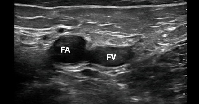
Switching to a linear probe and imaging at the common femoral artery has become our third step in our cardiac arrest ultrasound algorithm because this approach is faster and more reliable than subjectively feeling for a pulse. Ultrasound is more than twice as sensitive for the presence of a pulse than manual palpation.6 Using a linear probe to identify pulsatility of the femoral or carotid artery has been shown to be more accurate than manual palpation for pulses, and can almost always be done in less than five seconds.6 This modification removes the clinical sonographer from the chest, decreasing prolonged pauses in CPR. The simplest way to look for a pulse with ultrasound is to place a linear probe on the inguinal crease and identify the common femoral artery during ongoing compressions. The right femoral region is commonly selected because the first two steps of the CASA protocol are usually performed from the right side of the patient. The goal is to apply enough pressure to collapse the femoral vein, and look for pulsatility of the femoral artery (see Figure 2).

You Might Also Like
  • Introducing the CASA Exam: A New Protocol to Guide Point-Of-Care Ultrasound in Cardiac Arrest
  • Survival After Cardiac Arrest May Depend on Which EMS Agency Shows Up
  • Pediatric In-Hospital Cardiac Arrest: Lower Survival During Nights, Weekends
Explore This Issue
ACEP Now: Vol 42 – No 09 – September 2023

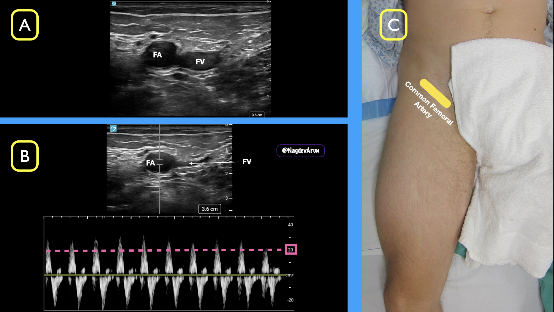
FIGURE 2: A) Compress the common femoral vein (FV) and visualize pulses in the common femoral artery (FA) B) Using spectral doppler, define a peak systolic velocity (PSV) >20 cm/sec C) Using a linear probe in the inguinal crease, locate the common femoral artery and vein. (Click to enlarge.)

In addition to using B-mode ultrasound to determine the presence or absence of a femoral pulse during a cardiac resuscitation, spectral or gated Doppler can be added at the same location to identify whether there is a perfusable rhythm. Work by Gaspari, et al., defined that some patients previously thought to have PEA are now recognized as having “pseudo-PEA,” where the heart is beating in an organized manner but a pulse cannot be manually palpated due to low cardiac output.7 This scenario raises the question of how to identify if sufficient cardiac perfusion is present that CPR is no longer needed. Recent studies show that applying gated Doppler to the femoral artery can identify patients who have achieved ROSC whether or not they have a palpable pulse.8 In practical terms, if femoral arterial pulsation is present on B-mode ultrasound during a pulse check, the next step is to use the spectral Doppler function to measure the peak systolic velocity, which may be used to define a perfusable pressure (or ROSC). This technique allows the clinician to determine the presence of a perfusable rhythm and may be even more sensitive than a rise in the end-tidal CO2, which was previously considered the first sign of ROSC.9

Pages: 1 2 3 4 | Single Page

Topics: Cardiac ArrestClinicalCPRImaging & UltrasoundPOCUSPoint-of-Care UltrasoundQuality & SafetyResuscitation

Related

  • Non-Invasive Positive Pressure Ventilation in the Emergency Department

    October 1, 2025 - 0 Comment
  • Emergency Department Management of Prehospital Tourniquets

    October 1, 2025 - 0 Comment
  • ACEP’s October 2025 Poll: How Often Do You Read Your Own X-Rays?

    September 30, 2025 - 0 Comment

Current Issue

ACEP Now: November 2025

Download PDF

Read More

No Responses to “How To Safely Incorporate Ultrasound Into Cardiac Arrest Resuscitation”

Leave a Reply Cancel Reply

Your email address will not be published. Required fields are marked *


*
*


Wiley
  • Home
  • About Us
  • Contact Us
  • Privacy
  • Terms of Use
  • Advertise
  • Cookie Preferences
Copyright © 2025 by John Wiley & Sons, Inc. All rights reserved, including rights for text and data mining and training of artificial technologies or similar technologies. ISSN 2333-2603