
Explore This Issue
ACEP Now: Vol 34 – No 08 – August 2015(Click for larger image)
Figure 2. Identify the patella and then slide the transducer cephalad until the approach ultrasound landmarks, including the patella, femur, quadriceps femoris tendon, and fat pad, are appreciated. The effusion will appear as an anechoic (black) space underneath the fat pad. (Green circle indicates ultrasound transducer directional marker.) Credit: Arun Nagdev
Ultrasound-Guided Knee Arthrocentesis
Materials:
- Sterile ultrasound sheath and gel
- 18g 1.5 needle attached to a 5–10 mL syringe
- Chlorhexidine
- Sterile drape
- Sterile gloves
- 30 g tuberculin syringe filled with 1–2% lidocaine or ethyl chloride spray
*Pictures are for educational purposes, and sterile precautions (sterile probe cover, gloves, drapes, etc.) should be used for all joint aspirations.
(Click for larger image)
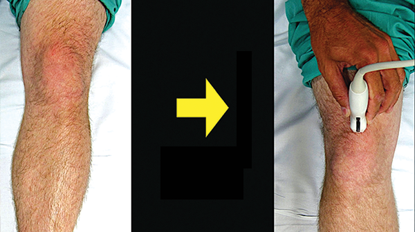
Figure 3. Rotate the probe marker to the patient’s right in order to visualize a transverse view of the prepatellar space with the effusion. Credit: Arun Nagdev
Joint aspiration:
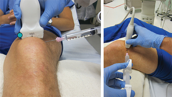
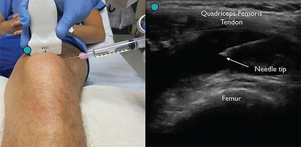
For ultrasound-guided arthrocentesis, we recommend a lateral to medial in-plane technique. With the linear probe in the prepatellar fossa, rotate the probe marker (clockwise) to the patient’s right to obtain a transverse view of the prepatellar space (see Figure 3). A large area around the suprapatellar space should be cleaned and draped in a sterile manner. The ultrasound transducer should be placed in a sterile sheath and then on the sterile field. The ultrasound system should be positioned opposite to the provider so the screen is in the direct line of sight. Using a 10 mL syringe attached to a standard 18g needle, enter the skin in plane and just lateral to the probe at a shallow angle (see Figure 4). The needle will traverse between the iliotibial band (superiorly) and vastus lateralis (inferiorly) without risk for vascular puncture. Clear needle visualization can be achieved by slowly advancing just under the transducer. Gentle aspiration of synovial fluid with needle tip being visualized within the fluid collection will confirm violation of the joint space.
(Click for larger image)
Figure 4. Enter the skin lateral to the probe using an 18g needle attached to a syringe for an in-plane arthrocentesis while visualizing the needle tip. Credit: Arun Nagdev
Summary
Point-of-care ultrasound can be a useful adjunct in the evaluation of the patient with a swollen, painful knee. Ultrasound can identify a suspected knee effusion as well as assist with arthrocentesis. A simplified in-plane technique can easily be incorporated into the evaluation of the patient with a suspected septic knee.
Pages: 1 2 3 | Single Page


One Response to “How to Perform Ultrasound-Guided Knee Arthrocentesis”
September 30, 2018
CassandraIs ultrasound injection good for ten knee after a keyhole surgery? I had this surgery for sometimes ago and now my left is severe with pain and decided to go for the ultrasound injection.. I had this injection on my shoulder and is good. I had it twice and never back again.So thinking of having one on my knee. Can you
Advise please thanks.