Explore This Issue
ACEP Now: Vol 39 – No 04 – April 2020Figure 2
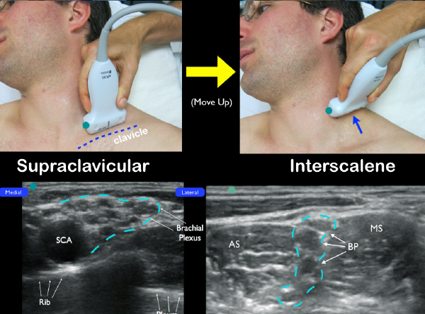
An alternative technique to locate the interscalane brachial plexus. Start with the transducer in the supraclavicular fossa. Locate the subclavian artery (SCA) in cross-section and note the brachial plexus just lateral (dotted blue line). Slide up the neck until the interscalene brachial plexus (BP) is located in between the anterior scalene (AS) and middle scalene (MS) muscles.
PHOTOS: Arun Nagdev
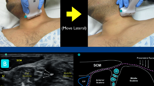
2) A fascial landmark to improve safety: Get anesthetic under the prevertebral fascia.
When performing ultrasound-guided femoral nerve blocks, we recommend depositing anesthetic under the fascia iliaca (lateral to the femoral nerve) to improve success. This target allows the clinician to enter far away from the nerve bundle, inject normal saline for hydrodissection, and then gently deposit anesthetic. This “stay away” approach has been our standard teaching technique (if possible) for all ultrasound-guided nerve blocks. Similarly, when performing the ultrasound-guided ISNB, the prevertebral fascia can act as an ideal sonographic landmark. This fascial plane lies on top of both the middle and anterior scalene muscles and allows the clinician a target that is far from the brachial plexus nerve roots (see Figures 3 and 4). With this ultrasound landmark in view, the clinician can come from a lateral to medial in-plane approach, then deposit anechoic normal saline (for hydrodissection) under the prevertebral fascia (far away from the interscalene brachial plexus). After getting under the fascial plane and visualizing fluid track down into the interscalene groove, anesthetic can safely be placed in the fluid-filled potential space (see Figures 5 and 6). In our opinion, placing the needle tip directly into the interscalene groove and between the nerve roots is not advisable, and it may lead to unwanted intraneural injections.
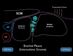
Figure 3
Schematic representation of the interscalene brachial plexus. Note the prevertebral fascia located above the middle and anterior scalene muscles.
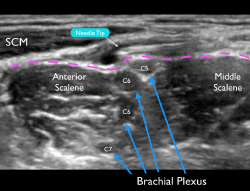
Figure 4
Ultrasound image of a failed block. Note that the needle tip is above the prevertebral fascia (pink dotted line). A small amount of anechoic anesthetic can be noted deposited above this important sonographic landmark.
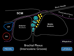
Figure 5
Schematic of how the needle tip should be placed just under the prevertebral fascia (and away from the roots of the brachial plexus).
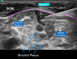
Figure 6
Ultrasound image of the needle tip under the prevertebral fascia (pink dotted line) with anechoic anesthetic deposited near the interscalene brachial plexus.
Pages: 1 2 3 | Single Page


One Response to “How to Perform Ultrasound-Guided Interscalene Nerve Blocks”
May 6, 2020
Vir SinghExcited to implement this in my practice and share this with my residency program