Logo

Log In Sign Up |  An official publication of: American College of Emergency Physicians
Navigation
  • Home
  • Multimedia
    • Podcasts
    • Videos
  • Clinical
    • Airway Managment
    • Case Reports
    • Critical Care
    • Guidelines
    • Imaging & Ultrasound
    • Pain & Palliative Care
    • Pediatrics
    • Resuscitation
    • Trauma & Injury
  • Resource Centers
    • mTBI Resource Center
  • Career
    • Practice Management
      • Benchmarking
      • Reimbursement & Coding
      • Care Team
      • Legal
      • Operations
      • Quality & Safety
    • Awards
    • Certification
    • Compensation
    • Early Career
    • Education
    • Leadership
    • Profiles
    • Retirement
    • Work-Life Balance
  • Columns
    • ACEP4U
    • Airway
    • Benchmarking
    • Brief19
    • By the Numbers
    • Coding Wizard
    • EM Cases
    • End of the Rainbow
    • Equity Equation
    • FACEPs in the Crowd
    • Forensic Facts
    • From the College
    • Images in EM
    • Kids Korner
    • Medicolegal Mind
    • Opinion
      • Break Room
      • New Spin
      • Pro-Con
    • Pearls From EM Literature
    • Policy Rx
    • Practice Changers
    • Problem Solvers
    • Residency Spotlight
    • Resident Voice
    • Skeptics’ Guide to Emergency Medicine
    • Sound Advice
    • Special OPs
    • Toxicology Q&A
    • WorldTravelERs
  • Resources
    • ACEP.org
    • ACEP Knowledge Quiz
    • Issue Archives
    • CME Now
    • Annual Scientific Assembly
      • ACEP14
      • ACEP15
      • ACEP16
      • ACEP17
      • ACEP18
      • ACEP19
    • Annals of Emergency Medicine
    • JACEP Open
    • Emergency Medicine Foundation
  • About
    • Our Mission
    • Medical Editor in Chief
    • Editorial Advisory Board
    • Awards
    • Authors
    • Article Submission
    • Contact Us
    • Advertise
    • Subscribe
    • Privacy Policy
    • Copyright Information

How To Perform an Ultrasound-Guided Clavipectoral Block

By Henry Ashworth, MD; Karen Lind, MD; David Martin, MD; Arun Nagdev, MD | on February 11, 2024 | 1 Comment
Sound Advice
  • Tweet
  • Click to email a link to a friend (Opens in new window) Email
Print-Friendly Version

FIGURE 2B: The goal is to visualize the needle tip until it touches the clavicle (red line). (Click to enlarge.)[/caption]

You Might Also Like
  • How To Perform an Ultrasound-Guided TAP Block for Appendicitis Pain
  • How to Perform Ultrasound-Guided Distal Sciatic Nerve Block in the Popliteal Fossa
  • Pain Control Using Ultrasound-Guided Superficial Cervical Plexus Block
Explore This Issue
ACEP Now: Vol 43 – No 02 – February 2024

Once the relevant anatomy is located, place a small skin wheal (2 to 3 ccs of anesthetic) at the inferior aspect of the transducer. Along with marking the entry point for the block, this will provide analgesic for the block.

3. Supplies and Anesthetic

The CPB block uses similar equipment as other ultrasound guided nerve blocks:

  • An ultrasound system with a high-frequency 15-6 MHz or 10-5 MHz linear transducer on musculoskeletal or soft-tissue setting
  • A blunt-tipped needle: preferably a 20- or 21-gauge regional-block needle
  • Appropriately sized syringe to draw local anesthetic; we recommend 20 mL
  • 10 mL sterile saline flushes for hydrodissection
  • Transparent adhesive ultrasound probe cover (Tegaderm or similar)
  • Antiseptic (e.g., alcohol, povidone-iodine, chlorhexidine)
  • Sterile ultrasound gel. If available, we recommend using ropivacaine (a concentration of 0.25 percent or less) as it has a longer half-life and a better safety profile than bupivacaine. Weight-based dosing should always be used to prevent local anesthetic systemic toxicity.

4. Performing Procedure

FIGURE 3: The ultrasound system screen needs to be in clear line of sight so that the clinician can see both needle and ultrasound

image. (Click to enlarge.)

Before performing the block, make sure the patient has consented, has intravenous access and is placed on a cardiac monitor.

The patient should be in a supine position. The ultrasound system should be placed so that the operator is able to visualize the point of needle entry, as well as the ultrasound screen in the same line of sight. We commonly place the ultrasound system contralateral to the clavicle that will be blocked (based on what is comfortable for the physician) (Figure 3).

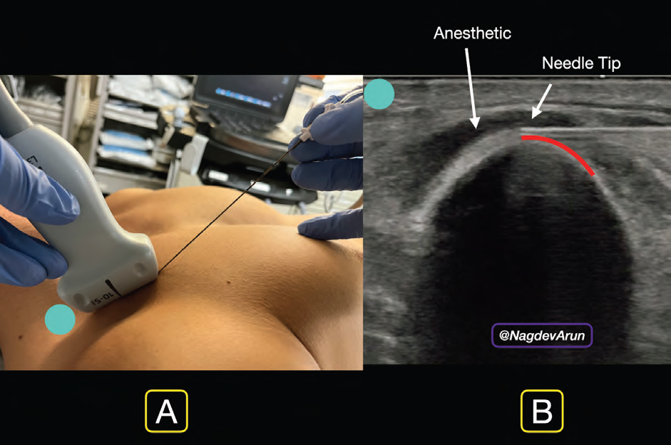
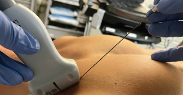
FIGURE 4A: In-plane needle entry.

FIGURE 4B: Needle tip visualized clearly with anesthetic deposited under the clavipectoral fascia. (Click to enlarge.)

Using an in-plane needle approach, enter the skin wheal with a block needle. Visualize the needle tip advancing through the pectoralis major to the inferior aspect of the clavicle (Figure 4A). Ensure continuous needle visualization until the block needle meets the clavicle; resistance may be felt as it goes through the clavipectoral fascia and touches the ostium of the clavicle. We recommend placing the needle at the 12 to 2 o’clock position of the clavicle (Figure 4B). Gently inject 1 to 2 cc of anesthetic to hydrodissect the clavipectoral fascia from the clavicle. The goal is to use the anechoic anesthetic to lift the pectoralis muscle away from the clavicle. This can be visualized with real-time ultrasound guidance. Once the clinician opens this potential space, gently place 3- to 5-cc aliquots, aspirating in between to ensure lack of inadvertent vascular puncture.

Pages: 1 2 3 4 | Single Page

Topics: clavipectoral nerve blocksClinicalNerve BlocksPain & Palliative CareUltrasound-Guided Nerve Block

Related

  • Non-Invasive Positive Pressure Ventilation in the Emergency Department

    October 1, 2025 - 0 Comment
  • Emergency Department Management of Prehospital Tourniquets

    October 1, 2025 - 0 Comment
  • ACEP’s October 2025 Poll: How Often Do You Read Your Own X-Rays?

    September 30, 2025 - 0 Comment

Current Issue

ACEP Now: November 2025

Download PDF

Read More

One Response to “How To Perform an Ultrasound-Guided Clavipectoral Block”

  1. February 28, 2024

    Doc_C Reply

    Why not simply a superficial cervical plexus block more on the caudal side to get the rami claviculares of the cervical plexus?

    Cheers

Leave a Reply Cancel Reply

Your email address will not be published. Required fields are marked *


*
*


Wiley
  • Home
  • About Us
  • Contact Us
  • Privacy
  • Terms of Use
  • Advertise
  • Cookie Preferences
Copyright © 2025 by John Wiley & Sons, Inc. All rights reserved, including rights for text and data mining and training of artificial technologies or similar technologies. ISSN 2333-2603