Logo

Log In Sign Up |  An official publication of: American College of Emergency Physicians
Navigation
  • Home
  • Multimedia
    • Podcasts
    • Videos
  • Clinical
    • Airway Managment
    • Case Reports
    • Critical Care
    • Guidelines
    • Imaging & Ultrasound
    • Pain & Palliative Care
    • Pediatrics
    • Resuscitation
    • Trauma & Injury
  • Resource Centers
    • mTBI Resource Center
  • Career
    • Practice Management
      • Benchmarking
      • Reimbursement & Coding
      • Care Team
      • Legal
      • Operations
      • Quality & Safety
    • Awards
    • Certification
    • Compensation
    • Early Career
    • Education
    • Leadership
    • Profiles
    • Retirement
    • Work-Life Balance
  • Columns
    • ACEP4U
    • Airway
    • Benchmarking
    • Brief19
    • By the Numbers
    • Coding Wizard
    • EM Cases
    • End of the Rainbow
    • Equity Equation
    • FACEPs in the Crowd
    • Forensic Facts
    • From the College
    • Images in EM
    • Kids Korner
    • Medicolegal Mind
    • Opinion
      • Break Room
      • New Spin
      • Pro-Con
    • Pearls From EM Literature
    • Policy Rx
    • Practice Changers
    • Problem Solvers
    • Residency Spotlight
    • Resident Voice
    • Skeptics’ Guide to Emergency Medicine
    • Sound Advice
    • Special OPs
    • Toxicology Q&A
    • WorldTravelERs
  • Resources
    • ACEP.org
    • ACEP Knowledge Quiz
    • Issue Archives
    • CME Now
    • Annual Scientific Assembly
      • ACEP14
      • ACEP15
      • ACEP16
      • ACEP17
      • ACEP18
      • ACEP19
    • Annals of Emergency Medicine
    • JACEP Open
    • Emergency Medicine Foundation
  • About
    • Our Mission
    • Medical Editor in Chief
    • Editorial Advisory Board
    • Awards
    • Authors
    • Article Submission
    • Contact Us
    • Advertise
    • Subscribe
    • Privacy Policy
    • Copyright Information

How To Perform an Ultrasound-Guided Clavipectoral Block

By Henry Ashworth, MD; Karen Lind, MD; David Martin, MD; Arun Nagdev, MD | on February 11, 2024 | 1 Comment
Sound Advice
  • Tweet
  • Click to email a link to a friend (Opens in new window) Email
Print-Friendly Version

The clavicle is one of the most commonly fractured bones in the human body. Every  year, approximately 332,000 people in the United States suffer from a clavicular fracture.1,2 In the emergency department (ED), the focus of treatment is pain control and immobilization unless there is a clear indication for surgery, such as open fractures, skin tenting, or neurovascular compromise.1,2 The mainstay of pain control in the ED has been either oral or intravenous non-steroidal anti-inflammatory drugs (NSAIDs) or opioids.2 However, in our experience these agents commonly do not offer optimal pain control in the acute setting, leading to repeat dosing and, often, prolonged ED stays.

You Might Also Like
  • How To Perform an Ultrasound-Guided TAP Block for Appendicitis Pain
  • How to Perform Ultrasound-Guided Distal Sciatic Nerve Block in the Popliteal Fossa
  • Pain Control Using Ultrasound-Guided Superficial Cervical Plexus Block
Explore This Issue
ACEP Now: Vol 43 – No 02 – February 2024

The ultrasound-guided clavipectoral plane block (CPB) is a newly described technique in the emergency medicine literature.3 The CPB is a simplified technique that can be easily integrated into clinical care by visualizing the deposition of anesthetic in the fascial plane around the clavicle.3,4 While the CPB was first described in the perioperative setting, it may be even more impactful in the ED setting after an acute injury.4-6

1. Anatomy

The clavicle is enwrapped by the clavipectoral fascia, which extends inferiorly from the clavicle, deep to the pectoralis major and superficial to the subclavius muscles. The innervation of the clavicle is poorly understood partly due to anatomical variations.7 The CPB circumvents this issue by directly blocking the terminal sensory nerve endings that innervate the clavicle.3

2. Ultrasound Survey Scan

FIGURE 1A: Ultrasound probe should be placed at the clavicle with the probe marker facing cephalad (toward the head).

FIGURE 1B: Ultrasound image with labeled relevant anatomy. (Click to enlarge.)

The clinician should note the fracture site (either via palpable landmark or ultrasound visualization). The initial survey scan should be either just lateral or medial to this site.

The anatomy for the CPB is visualized by placing a linear transducer in a cranial-caudal direction at the midclavicular line with the probe marker towards the patient’s head (Figure 1A). In this position, the pectoralis major and subclavius muscle are located just below the clavicle (Figure 1A). The goal of this simple block is to place anesthetic gently under the fascial plane that defines the border of the pectoralis muscle and the clavicle (Figure 2A, 2B). In our experience, placing anesthetic near the top of the clavicle will separate this potential space and allow for optimal fracture analgesia.

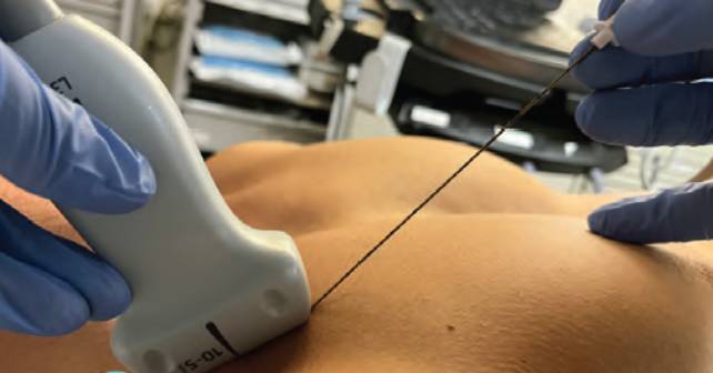
FIGURE 2A: Ultrasound probe placed in a sagittal plane with the probe marker cephalad, in-plane needle technique.

Pages: 1 2 3 4 | Single Page

Topics: clavipectoral nerve blocksClinicalNerve BlocksPain & Palliative CareUltrasound-Guided Nerve Block

Related

  • Non-Invasive Positive Pressure Ventilation in the Emergency Department

    October 1, 2025 - 0 Comment
  • Emergency Department Management of Prehospital Tourniquets

    October 1, 2025 - 0 Comment
  • ACEP’s October 2025 Poll: How Often Do You Read Your Own X-Rays?

    September 30, 2025 - 0 Comment

Current Issue

ACEP Now: November 2025

Download PDF

Read More

One Response to “How To Perform an Ultrasound-Guided Clavipectoral Block”

  1. February 28, 2024

    Doc_C Reply

    Why not simply a superficial cervical plexus block more on the caudal side to get the rami claviculares of the cervical plexus?

    Cheers

Leave a Reply Cancel Reply

Your email address will not be published. Required fields are marked *


*
*


Wiley
  • Home
  • About Us
  • Contact Us
  • Privacy
  • Terms of Use
  • Advertise
  • Cookie Preferences
Copyright © 2025 by John Wiley & Sons, Inc. All rights reserved, including rights for text and data mining and training of artificial technologies or similar technologies. ISSN 2333-2603