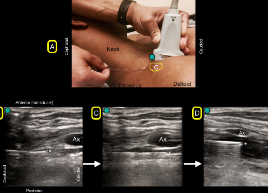
Logo

Log In Sign Up |  An official publication of: American College of Emergency Physicians
Navigation
  • Home
  • Multimedia
    • Podcasts
    • Videos
  • Clinical
    • Airway Managment
    • Case Reports
    • Critical Care
    • Guidelines
    • Imaging & Ultrasound
    • Pain & Palliative Care
    • Pediatrics
    • Resuscitation
    • Trauma & Injury
  • Resource Centers
    • mTBI Resource Center
  • Career
    • Practice Management
      • Benchmarking
      • Reimbursement & Coding
      • Care Team
      • Legal
      • Operations
      • Quality & Safety
    • Awards
    • Certification
    • Compensation
    • Early Career
    • Education
    • Leadership
    • Profiles
    • Retirement
    • Work-Life Balance
  • Columns
    • ACEP4U
    • Airway
    • Benchmarking
    • Brief19
    • By the Numbers
    • Coding Wizard
    • EM Cases
    • End of the Rainbow
    • Equity Equation
    • FACEPs in the Crowd
    • Forensic Facts
    • From the College
    • Images in EM
    • Kids Korner
    • Medicolegal Mind
    • Opinion
      • Break Room
      • New Spin
      • Pro-Con
    • Pearls From EM Literature
    • Policy Rx
    • Practice Changers
    • Problem Solvers
    • Residency Spotlight
    • Resident Voice
    • Skeptics’ Guide to Emergency Medicine
    • Sound Advice
    • Special OPs
    • Toxicology Q&A
    • WorldTravelERs
  • Resources
    • ACEP.org
    • ACEP Knowledge Quiz
    • Issue Archives
    • CME Now
    • Annual Scientific Assembly
      • ACEP14
      • ACEP15
      • ACEP16
      • ACEP17
      • ACEP18
      • ACEP19
    • Annals of Emergency Medicine
    • JACEP Open
    • Emergency Medicine Foundation
  • About
    • Our Mission
    • Medical Editor in Chief
    • Editorial Advisory Board
    • Awards
    • Authors
    • Article Submission
    • Contact Us
    • Advertise
    • Subscribe
    • Privacy Policy
    • Copyright Information

How to Effectively Block an Acutely Fractured Distal Radius

By Josh Luftig, PA; Daniel Mantuani, MD; and Arun Nagdev, MD | on October 16, 2018 | 2 Comments
Sound Advice
  • Tweet
  • Click to email a link to a friend (Opens in new window) Email
Print-Friendly Version
Figure 4A: A flat needle angle will be used to traverse under the clavicle (C). 4B: The bock needle will have to pass under the clavicle and will not be visualized. Note the needle tip as it emerges from under the clavicle and can be easily seen because of its flat angle. 4C: The block needle is advanced until placed just under the axillary artery (Ax). 4D: Anechoic anesthetic fluid is injected under the axillary artery (Ax) for a successful block.

You Might Also Like
  • Pain Control Using Ultrasound-Guided Superficial Cervical Plexus Block
  • How to Perform Ultrasound-Guided Distal Sciatic Nerve Block in the Popliteal Fossa
  • Ultrasound-Guided Posterior Tibial Nerve Block
Explore This Issue
ACEP Now: Vol 37 – No 10 – October 2018

Figure 4A: A flat needle angle will be used to traverse under the clavicle (C). 4B: The bock needle will have to pass under the clavicle and will not be visualized. Note the needle tip as it emerges from under the clavicle and can be easily seen because of its flat angle. 4C: The block needle is advanced until placed just under the axillary artery (Ax). 4D: Anechoic anesthetic fluid is injected under the axillary artery (Ax) for a successful block.
Credit: Luftig, Mantuani, Nagdev

Place a local anesthetic skin wheal at the insertion site using a 25–27g needle, then insert the block needle through the skin wheal and advance the needle beneath the clavicle toward the ultrasound beam at an angle parallel to the gurney (see Figure 4A). The patient should be prompted to alert the operator of any paresthesia in the shoulder while the needle advances, as the suprascapular nerve lies in the vicinity; this is a very uncommon occurrence. If the patient reports paresthesia of the shoulder, the needle should be withdrawn, and a slightly different needle path should be used. Also, when passing through the blind zone created by the clavicle, the needle should never be angled posteriorly, as this increases the risk of pneumothorax. However, the entire needle can be posteriorly deflected by applying pressure to the shaft at the insertion site while maintaining the same flat angle of approach, similar to the method used to get past the clavicle for a blind subclavian central line.

After insertion through the approximately 3 cm blind zone, use subtle needle and transducer adjustments to locate the clearly visualized needle emerging from beneath the clavicle (see Figure 4B). Continue advancing with in-plane ultrasound guidance toward the target location just posterior to the axillary artery, typically at or slightly past the six o’clock position relative to the artery (see Figure 4C). Aspirate to check for inadvertent vascular puncture and then inject small aliquots of normal saline. Anechoic anesthetic fluid should be seen spreading just posterior to the axillary artery and deflecting the artery toward the probe, confirming needle tip location within the axillary sheath (see Figure 4D). The sheath surrounds both the brachial plexus and the axillary artery. Thus, fluid injected into the sheath bathes the plexus. It is not necessary to visualize the nerves of the plexus. Once satisfied with the needle position, gradually inject local anesthetic until a total of 35–40 mL is deposited within the sheath. Total needling time is usually less than five minutes, and dense arm anesthesia develops within 30.

Pages: 1 2 3 4 5 | Single Page

Topics: Emergency UltrasoundPain & Palliative CareUltrasoundUltrasound-Guided Nerve Block

Related

  • Case Report: Rare Pulmonary Embolism After Routine PIVC Insertion

    September 22, 2025 - 1 Comment
  • 10 Essentials for Your Emergency Department Fanny Pack

    June 17, 2025 - 0 Comment
  • Case Report: Rapid Diagnosis of Acute Aortic Dissection with POCUS

    June 11, 2025 - 1 Comment

Current Issue

ACEP Now: November 2025

Download PDF

Read More

2 Responses to “How to Effectively Block an Acutely Fractured Distal Radius”

  1. November 4, 2018

    Mark Bailey Reply

    Nice if the ED MD can have a specialist come down. Too much time wasted for a real ED.

  2. November 4, 2018

    Michael Anthony Macias Reply

    Love this block and looking forward to incorporating into my practice though probably should reserve for more complex injuries as majority of distal radius fractures are easily reduced comfortably with a simple hematoma block.

Leave a Reply Cancel Reply

Your email address will not be published. Required fields are marked *


*
*


Wiley
  • Home
  • About Us
  • Contact Us
  • Privacy
  • Terms of Use
  • Advertise
  • Cookie Preferences
Copyright © 2025 by John Wiley & Sons, Inc. All rights reserved, including rights for text and data mining and training of artificial technologies or similar technologies. ISSN 2333-2603