A 40-year-old woman presented to our emergency department complaining of left hemiparesis and headache. She was awake and oriented, and her vitals were unremarkable except for a blood pressure of 180/100 without a history of hypertension. Past history and medications were negative. No trauma was reported. The optic nerve sheath diameter (ONSD) was measured and showed increased diameter of 0.47 cm bilaterally, demonstrating increased intracranial pressure (see Figure 1). Her level of consciousness decreased dramatically within 30 minutes, and she developed a right-sided gaze. She was intubated and underwent a brain CT showing intracranial hemorrhage in the right basal ganglia with midline shift and intraventricular hemorrhage (see Figure 2). She received a phenytoin loading dose and was taken emergently to the operating room. Fortunately, she survived with an acceptable neurologic outcome.
Explore This Issue
ACEP Now: Vol 36 – No 06 – June 2017ONSD can predict increased intracranial pressure effectively. The exact measurement can be achieved by frequent practice. Thus, this approach is being described as an operator-dependent procedure.
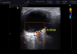
Figure 1: The optic nerve sheath diameter was measured at 0.47 cm, demonstrating an increased intracranial process in our patient.
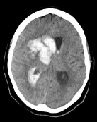
Figure 2: Brain computed tomography showed intracranial hemorrhage of the right basal ganglia as well as an intraventricular hemorrhage and a significant midline shift.
Optic Nerve Sheath Diameter
There is no doubt that ultrasonography is a valuable way to evaluate ONSD to determine intracranial pressure, considering that the brain subarachnoid space is continuous with the optic nerve sheath and the pressure should be transmitted.1–4 However, the way to measure the diameter has yet to be determined. Optic nerve color Doppler ultrasonography may clarify this measure and change what was previously approved to evaluate the ONSD. Copetti et al noted that the former measures were not compatible with the real anatomy of the optic nerve and proposed a new measurement that seems to be more reliable.1 There are two points to be considered:
- According to the route of optic nerve approaching the globe, the oblique hypoechoic shadow running medially represents the exact measure of the optic nerve (see Figure 3).
- Regarding color Doppler ultrasonography of the central retinal artery, which runs within the dural optic nerve sheath to the globe, the new image compatible with the oblique hypoechoic shadow seems more characteristic of the optic nerve.

Figure 3: Ocular ultrasound with a 12 MHz linear probe in a normal subject shows a 35 mm optic nerve diameter, identified with color Doppler.
Pages: 1 2 3 | Single Page


No Responses to “Emergency Ocular Ultrasonography to Measure Optic Nerve Sheath Diameter”