Logo

Log In Sign Up |  An official publication of: American College of Emergency Physicians
Navigation
  • Home
  • Multimedia
    • Podcasts
    • Videos
  • Clinical
    • Airway Managment
    • Case Reports
    • Critical Care
    • Guidelines
    • Imaging & Ultrasound
    • Pain & Palliative Care
    • Pediatrics
    • Resuscitation
    • Trauma & Injury
  • Resource Centers
    • mTBI Resource Center
  • Career
    • Practice Management
      • Benchmarking
      • Reimbursement & Coding
      • Care Team
      • Legal
      • Operations
      • Quality & Safety
    • Awards
    • Certification
    • Compensation
    • Early Career
    • Education
    • Leadership
    • Profiles
    • Retirement
    • Work-Life Balance
  • Columns
    • ACEP4U
    • Airway
    • Benchmarking
    • Brief19
    • By the Numbers
    • Coding Wizard
    • EM Cases
    • End of the Rainbow
    • Equity Equation
    • FACEPs in the Crowd
    • Forensic Facts
    • From the College
    • Images in EM
    • Kids Korner
    • Medicolegal Mind
    • Opinion
      • Break Room
      • New Spin
      • Pro-Con
    • Pearls From EM Literature
    • Policy Rx
    • Practice Changers
    • Problem Solvers
    • Residency Spotlight
    • Resident Voice
    • Skeptics’ Guide to Emergency Medicine
    • Sound Advice
    • Special OPs
    • Toxicology Q&A
    • WorldTravelERs
  • Resources
    • ACEP.org
    • ACEP Knowledge Quiz
    • Issue Archives
    • CME Now
    • Annual Scientific Assembly
      • ACEP14
      • ACEP15
      • ACEP16
      • ACEP17
      • ACEP18
      • ACEP19
    • Annals of Emergency Medicine
    • JACEP Open
    • Emergency Medicine Foundation
  • About
    • Our Mission
    • Medical Editor in Chief
    • Editorial Advisory Board
    • Awards
    • Authors
    • Article Submission
    • Contact Us
    • Advertise
    • Subscribe
    • Privacy Policy
    • Copyright Information

Case Report: The Ice Pack Test

By Sam Cray, DO; Austin Bennett, DO | on August 8, 2024 | 0 Comment
Case Report
  • Tweet
  • Click to email a link to a friend (Opens in new window) Email
Print-Friendly Version

You Might Also Like
  • Case Report Provides Tips for Diagnosing the Rare Tolosa-Hunt Syndrome
  • A Case Report on Tetanus
  • Case Report: Toxic Hypoglycemic Syndrome
Explore This Issue
ACEP Now: Vol 43 – No 08 – August 2024

IMAGE 1: Forward gaze on presentation.

This patient is a 52-year-old male with past medical history of hypertension and hyperlipidemia who presented to the emergency department (ED) with approximately one month of progressive bilateral ptosis and one week of progressive bilateral diplopia and photophobia. The patient reported that these symptoms were more noticeable later in the day and worsened with reading and typing. The patient’s gaze disturbance was first noted by his wife, and he agreed to be evaluated by an optometrist. The patient was seen and instructed to report to the ED for concern of neuromuscular or intracranial pathology. After an unremarkable initial workup at an outside ED, which included head CT/CTA, blood chemistry, chest radiograph, and ECG, the patient was transferred to our tertiary care center for further neurological evaluation.

IMAGE 2: Upward gaze on presentation, note frontalis muscle compensation to lift eyelids.

The patient’s vital signs were within normal limits aside from mildly elevated blood pressure. Review of systems was positive for headache, diplopia, and photophobia. He denied recent fever, seizures, vertigo, limb weakness, stiffness, or pain. Reassuringly, the patient denied having any respiratory complaints. On physical exam, the patient exhibited bilateral ptosis (see Image 1). Extraocular range of motion was intact, but he had some difficulty with vertical gaze, which was easily fatigable and accompanied with noticeable compensation by the frontalis muscle (see Image 2). Further ocular testing revealed positive accommodation but marked photophobia. This prevented pupillary light reflex testing because the patient squeezed his eyelids shut to bright light. He was unable to complete visual field testing due to diplopia. The patient’s neck exhibited full active and passive range of motion with no tenderness or meningismus.

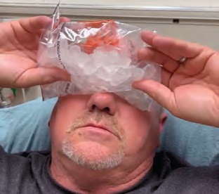
IMAGE 3: Administering the ice pack test.

The remainder of physical and neurological exams were unremarkable.

Diagnosis and Initial Treatment

The decision was then made to test for myasthenia gravis (MG) by application of an ice pack. A plastic bag was filled with ice, and the patient was instructed to hold this over his eyes for two minutes (Image 3). The ice pack was then removed, and both patient and examiners observed noticeable improvement. The patient’s ptosis was absent (Image 4). Extraocular range of motion was normal (Image 5), and the patient reported resolution of diplopia and photophobia. Pupillary light reflex was assessed and found to be normal. This effect lasted for approximately 90 seconds before ptosis reappeared. The test was conducted twice, approximately 15 minutes apart, with identical results, and the diagnosis of MG was made.

Pages: 1 2 3 4 | Single Page

Topics: case reportCase ReportsClinicalDiplopiamyasthenia gravisneuromuscularphotophobiaptosis

Related

  • Non-Invasive Positive Pressure Ventilation in the Emergency Department

    October 1, 2025 - 0 Comment
  • Emergency Department Management of Prehospital Tourniquets

    October 1, 2025 - 0 Comment
  • ACEP’s October 2025 Poll: How Often Do You Read Your Own X-Rays?

    September 30, 2025 - 0 Comment

Current Issue

ACEP Now: November 2025

Download PDF

Read More

No Responses to “Case Report: The Ice Pack Test”

Leave a Reply Cancel Reply

Your email address will not be published. Required fields are marked *


*
*


Wiley
  • Home
  • About Us
  • Contact Us
  • Privacy
  • Terms of Use
  • Advertise
  • Cookie Preferences
Copyright © 2025 by John Wiley & Sons, Inc. All rights reserved, including rights for text and data mining and training of artificial technologies or similar technologies. ISSN 2333-2603