Upper extremity DVT has traditionally been associated with central venous catheters, pacemakers, thoracic outlet syndrome, or malignancy. However, emerging data show that even peripheral interventions may cause endothelial injury sufficient to provoke thrombus formation, particularly when compounded by systemic risk factors such as infection, dehydration, and prior venous damage.2,4
Explore This Issue
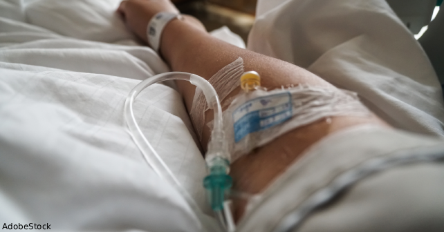
ACEP Now: October 2025 (Digital)The rate of PE occurrence in patients with UE-DVT is rarely reported. Additionally, as this patient had prior thrombophlebitis in the same location, it is plausible that local scarring and valvular dysfunction promoted stasis and increased the thrombogenic potential.3
Environmental stressors in the field like dehydration, high-altitude physiology, and limited diagnostic resources may amplify the risk for thrombosis.5 The gastroenteritis episode may have induced a transient hypercoagulable state through hemoconcentration and inflammatory cytokine release.6
Early identification of SVT, particularly in high-risk individuals, should prompt duplex ultrasonography to evaluate for contiguous DVT. Management of UE-DVT mirrors that of lower extremity DVT, including prompt initiation of anticoagulation. Guidelines from the American College of Chest Physicians support anticoagulation for symptomatic UE-DVT for at least three months.5
Conclusion
This case underscores the potential for a seemingly routine procedure like PIVC insertion to result in serious thromboembolic complications, particularly within the unique context of a deployed military environment. The progression from superficial thrombophlebitis to UE-DVT and ultimately to PE highlights a continuum of vascular injury that may be underrecognized, especially in patients lacking classic risk factors such as malignancy or central venous catheters.
The austere conditions of deployment, limited diagnostic capabilities, environmental stressors like dehydration and high altitude, and delayed access to advanced care can all exacerbate thrombotic risk. This patient’s prior history of venous injury, combined with a transient hypercoagulable state because of acute gastroenteritis, created a confluence of factors that contributed to thrombus formation and embolic progression. Emergency and military medicine physicians must be particularly vigilant for such complications in deployed settings, where early signs of vascular inflammation may be easily overlooked or attributed to benign causes.
This case emphasizes the importance of thorough assessment of intravenous access sites, particularly in patients with known prior vein injury or systemic stressors. Physicians should maintain a high index of suspicion for thrombotic events in patients presenting with pain, swelling, or palpable cords after PIVC insertion. When available, point-of-care ultrasound can facilitate early diagnosis and guide prompt anticoagulation, potentially averting life-threatening sequelae such as PE.
Although existing guidelines such as those from the American College of Chest Physicians support anticoagulation for symptomatic UE-DVT, they often focus on patients with central venous catheters or malignancy.5 As the operational and civilian medical communities continue to report similar cases, there is a need to better characterize thrombotic risk associated with peripheral IVs, especially in high-risk environments like combat zones.
Pages: 1 2 3 | Single Page


One Response to “Case Report: Rare Pulmonary Embolism After Routine PIVC Insertion”
September 28, 2025
Scott MankowitzInteresting story. Please stop creating new abbreviations and placing them in the title of the article.