1. The “Dissociated” Inflammatory Markers
The most striking feature here was the discordance between the CRP (2.5 mg/L) and the Procalcitonin (2.24 ng/mL).
• The Trap: Relying solely on the normal CRP led to the initial exclusion of osteomyelitis. In approximately 2–5% of AHO cases, the CRP can be normal on admission, particularly if the organism is less virulent (e.g., Kingella—though not the case here) or in early/walled-off S. aureus infections.
• The Clue: The Procalcitonin was the “truth-teller.” A PCT >2.0 in a pediatric patient without a clear source is highly specific for invasive bacterial infection (bacteremia/sepsis) and should trigger a hunt for the source regardless of the CRP.
2. The “Phantom” Limp
The history of a limp that “improved” and a normal gait in the ED is a common confounder in pediatric osteomyelitis.
• Children often guard intermittently.
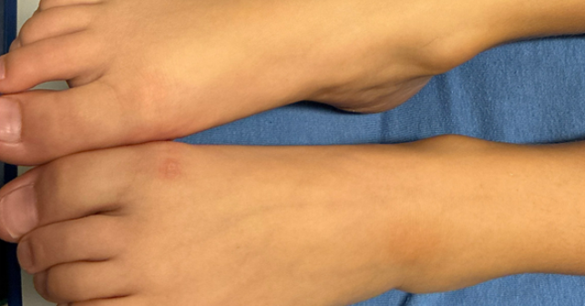
• The “lesion” on the ankle (initially thought to be the result of the dance shoe) was likely the cause (portal of entry) or a manifestation (septic embolus/Janeway lesion) of the intravascular infection.
3. T1DM as a Risk Factor (co-morbidities, co-morbidities, co-morbidities – ALWAYS be super suspect of more advanced or unusual disease)
Even with a normal glucose (81 mg/dL) and no DKA, the patient’s Type 1 Diabetes history is relevant.
• It places the patient at higher risk for invasive S. aureus infections (via pump sites/CGM or altered neutrophil function).
• It may also blunt the typical febrile/inflammatory response, contributing to the confusing presentation.
I have yet to see such a subtle cellulitis in someone mounting a fever of 104F. With that fever, there would be a correspondingly large area of typical indurated, very erythematous, tender, warm skin.
A subtle presentation on the derm exam combined with a fever of 104F does not, in my experience, align with a diagnosis of cellulitis.


3 Responses to “Case Report: Five Days of Fever”
December 21, 2025
Gabe WilsonA few learning points:
1. The “Dissociated” Inflammatory Markers
The most striking feature here was the discordance between the CRP (2.5 mg/L) and the Procalcitonin (2.24 ng/mL).
• The Trap: Relying solely on the normal CRP led to the initial exclusion of osteomyelitis. In approximately 2–5% of AHO cases, the CRP can be normal on admission, particularly if the organism is less virulent (e.g., Kingella—though not the case here) or in early/walled-off S. aureus infections.
• The Clue: The Procalcitonin was the “truth-teller.” A PCT >2.0 in a pediatric patient without a clear source is highly specific for invasive bacterial infection (bacteremia/sepsis) and should trigger a hunt for the source regardless of the CRP.
2. The “Phantom” Limp
The history of a limp that “improved” and a normal gait in the ED is a common confounder in pediatric osteomyelitis.
• Children often guard intermittently.
• The “lesion” on the ankle (initially thought to be the result of the dance shoe) was likely the cause (portal of entry) or a manifestation (septic embolus/Janeway lesion) of the intravascular infection.
3. T1DM as a Risk Factor (co-morbidities, co-morbidities, co-morbidities – ALWAYS be super suspect of more advanced or unusual disease)
Even with a normal glucose (81 mg/dL) and no DKA, the patient’s Type 1 Diabetes history is relevant.
• It places the patient at higher risk for invasive S. aureus infections (via pump sites/CGM or altered neutrophil function).
• It may also blunt the typical febrile/inflammatory response, contributing to the confusing presentation.
December 21, 2025
Gabe WilsonOne additional point:
DIAGNOSTIC INCONGRUITY/ANCHORING BIAS
I have yet to see such a subtle cellulitis in someone mounting a fever of 104F. With that fever, there would be a correspondingly large area of typical indurated, very erythematous, tender, warm skin.
A subtle presentation on the derm exam combined with a fever of 104F does not, in my experience, align with a diagnosis of cellulitis.
December 22, 2025
STEPHEN J. VAN CLEAVELimping is not common with cellulitis. So with joint pain in a diabetic child, you need CT or MRI of the affected joint, not plain X-ray.