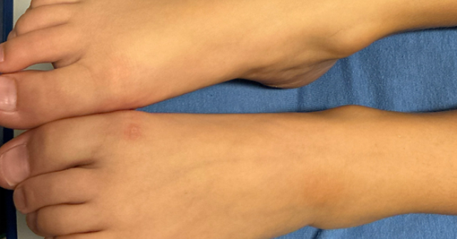

Our patient was misdiagnosed initially but, fortunately, a positive blood culture helped identify the diagnosis, and the patient was treated appropriately prior to development of permanent sequelae. The diagnosis of osteomyelitis is often difficult as it presents subclinically in the early stages and is relatively rare. Fever and pain are the most common presenting symptoms of osteomyelitis and can manifest as difficulty bearing weight or reduced use of the affected limb in pediatric patients.2 In our case the patient did have fever and a limp which was presumed secondary to sprain from the preceding dance lesson. A minimal skin abnormality consistent with inflammation was present, likely indicating the patient had an indolent infection. The use of inflammatory markers can guide the treatment plan, and an X-ray might show evidence of osteomyelitis. 3 Those were inconclusive on our patient and an MRI on initial presentation would have been ideal for diagnosis.3
The prevalence of osteomyelitis in children within developed nations is 13 per 100,000, however it is higher among those with diabetes.4,5 Research evaluating the intersectionality of type 1 diabetes mellitus and osteomyelitis in pediatric patients is extremely limited, therefore calculating an exact prevalence rate is difficult. It has been determined that patients with diabetes have a 1.5-4.5 times greater risk of infection than the general population.5 This makes the diagnosis a pertinent consideration when a diabetic patient presents with fever from an unclear source.
 Dr. Khalil Mroue is a clinical assistant professor at Burrell College of Osteopathic Medicine and at Hyatt Clinical Education. He currently works in emergency medicine at the Tucson Medical Center.
 Dominic Barandica is a fourth-year medical student at the Autonomous University of Guadalajara.
References
- Walter N, Bärtl S, Alt V, Rupp M. The epidemiology of osteomyelitis in children. Children (Basel). 2021;8(11):1000. doi:10.3390/children8111000.
- Woods CR, Bradley JS, Chatterjee A, et al. Clinical practice guideline by the Pediatric Infectious Diseases Society and the Infectious Diseases Society of America: 2021 guideline on diagnosis and management of acute hematogenous osteomyelitis in pediatrics. J Pediatric Infect Dis Soc. 2021;10(8):801-844. doi:10.1093/jpids/piab027.
- Hackenberg RK, Schmitt-Sánchez F, Endler C, et al. Value of Diagnostic Tools in the Diagnosis of Osteomyelitis: Pilot Study to Establish an Osteomyelitis Score. J Clin Med. 2023;12(9):3057. Published April 23, 2023.. doi:10.3390/jcm12093057.
- Momodu II, Savaliya V. Osteomyelitis. In: StatPearls. StatPearls Publishing; 2023. Accessed September 19, 2025. https://www.ncbi.nlm.nih.gov/books/NBK532250/.
- Holt RIG, Cockram CS, Ma RCW, Luk AOY. Diabetes and infection: Review of the epidemiology, mechanisms and principles of treatment. Diabetologia. 2024;67:1168-1180. doi:10.1007/s00125-024-06102-x.
Pages: 1 2 3 | Single Page


3 Responses to “Case Report: Five Days of Fever”
December 21, 2025
Gabe WilsonA few learning points:
1. The “Dissociated” Inflammatory Markers
The most striking feature here was the discordance between the CRP (2.5 mg/L) and the Procalcitonin (2.24 ng/mL).
• The Trap: Relying solely on the normal CRP led to the initial exclusion of osteomyelitis. In approximately 2–5% of AHO cases, the CRP can be normal on admission, particularly if the organism is less virulent (e.g., Kingella—though not the case here) or in early/walled-off S. aureus infections.
• The Clue: The Procalcitonin was the “truth-teller.” A PCT >2.0 in a pediatric patient without a clear source is highly specific for invasive bacterial infection (bacteremia/sepsis) and should trigger a hunt for the source regardless of the CRP.
2. The “Phantom” Limp
The history of a limp that “improved” and a normal gait in the ED is a common confounder in pediatric osteomyelitis.
• Children often guard intermittently.
• The “lesion” on the ankle (initially thought to be the result of the dance shoe) was likely the cause (portal of entry) or a manifestation (septic embolus/Janeway lesion) of the intravascular infection.
3. T1DM as a Risk Factor (co-morbidities, co-morbidities, co-morbidities – ALWAYS be super suspect of more advanced or unusual disease)
Even with a normal glucose (81 mg/dL) and no DKA, the patient’s Type 1 Diabetes history is relevant.
• It places the patient at higher risk for invasive S. aureus infections (via pump sites/CGM or altered neutrophil function).
• It may also blunt the typical febrile/inflammatory response, contributing to the confusing presentation.
December 21, 2025
Gabe WilsonOne additional point:
DIAGNOSTIC INCONGRUITY/ANCHORING BIAS
I have yet to see such a subtle cellulitis in someone mounting a fever of 104F. With that fever, there would be a correspondingly large area of typical indurated, very erythematous, tender, warm skin.
A subtle presentation on the derm exam combined with a fever of 104F does not, in my experience, align with a diagnosis of cellulitis.
December 22, 2025
STEPHEN J. VAN CLEAVELimping is not common with cellulitis. So with joint pain in a diabetic child, you need CT or MRI of the affected joint, not plain X-ray.