Logo

Log In Sign Up |  An official publication of: American College of Emergency Physicians
Navigation
  • Home
  • Multimedia
    • Podcasts
    • Videos
  • Clinical
    • Airway Managment
    • Case Reports
    • Critical Care
    • Guidelines
    • Imaging & Ultrasound
    • Pain & Palliative Care
    • Pediatrics
    • Resuscitation
    • Trauma & Injury
  • Resource Centers
    • mTBI Resource Center
  • Career
    • Practice Management
      • Benchmarking
      • Reimbursement & Coding
      • Care Team
      • Legal
      • Operations
      • Quality & Safety
    • Awards
    • Certification
    • Compensation
    • Early Career
    • Education
    • Leadership
    • Profiles
    • Retirement
    • Work-Life Balance
  • Columns
    • ACEP4U
    • Airway
    • Benchmarking
    • Brief19
    • By the Numbers
    • Coding Wizard
    • EM Cases
    • End of the Rainbow
    • Equity Equation
    • FACEPs in the Crowd
    • Forensic Facts
    • From the College
    • Images in EM
    • Kids Korner
    • Medicolegal Mind
    • Opinion
      • Break Room
      • New Spin
      • Pro-Con
    • Pearls From EM Literature
    • Policy Rx
    • Practice Changers
    • Problem Solvers
    • Residency Spotlight
    • Resident Voice
    • Skeptics’ Guide to Emergency Medicine
    • Sound Advice
    • Special OPs
    • Toxicology Q&A
    • WorldTravelERs
  • Resources
    • ACEP.org
    • ACEP Knowledge Quiz
    • Issue Archives
    • CME Now
    • Annual Scientific Assembly
      • ACEP14
      • ACEP15
      • ACEP16
      • ACEP17
      • ACEP18
      • ACEP19
    • Annals of Emergency Medicine
    • JACEP Open
    • Emergency Medicine Foundation
  • About
    • Our Mission
    • Medical Editor in Chief
    • Editorial Advisory Board
    • Awards
    • Authors
    • Article Submission
    • Contact Us
    • Advertise
    • Subscribe
    • Privacy Policy
    • Copyright Information

Case Report: Five Days of Fever

By Khalil Mroue, MD; Dominic Barandica | on December 16, 2025 | 3 Comments
Case Report
  • Tweet
  • Click to email a link to a friend (Opens in new window) Email
Print-Friendly Version

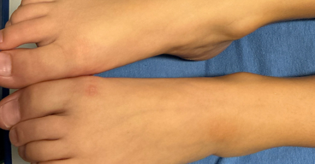
A 10-year-old boy with a history of type 1 diabetes mellitus presented to the emergency department (ED) with fevers for five days. Symptoms began with temperatures as high as 104⁰F with ketones in the urine noted on a home test. The patient did not have any associated symptoms. His mom stated that he had a limp that began after a dance rehearsal a few days ago, though no trauma was reported, and had since improved. The patient’s vaccination record was up to date. On exam, the patient was noted to have a slightly red, minimally tender, flat lesion of the left ankle which was not warm to touch (Image 1). His gait was normal. He had a fever but otherwise the rest of the vital signs were normal. Laboratory tests and an X-ray of the ankle were obtained. Results included: WBC of 6.4 x 10³ /µL, CRP of 2.5 mg/L (ref <4.90 mg/L), and procalcitonin of 2.24 ng/mL (ref 0.00-0.50 ng/mL). The chemistry panel was normal with blood glucose 81 mg/dL and no evidence of metabolic acidosis. The ankle X-ray showed soft tissue swelling without acute osseous abnormality. Blood cultures were sent. The patient was presumed to have cellulitis given skin discoloration and swelling on X-ray. He was given ceftriaxone in the ED but developed a rash,and was discharged on doxycycline.

You Might Also Like
  • Tips for Catching Commonly Missed Ankle Injuries
  • Tips for Managing Weber B Ankle Fractures
  • Rat-Bite Fever’s Non-Specific Symptoms Make Patient History Important for Diagnosis
Explore This Issue
ACEP Now: December 2025 (Digital)

Image 1. Note the subtle ankle rash on left anterolateral ankle. (Click to enlarge.)

The following day, blood cultures were positive for Staphylococcus aureus (MSSA). The patient was called to return and subsequently admitted for bacteremia. An MRI of the ankle was obtained, which revealed osteomyelitis with a small amount (1 mL) of fluid along the anterior distal tibia. Patient was subsequently taken to the operating room with orthopedics for drainage and decompression. Following this, he continued to improve with IV antibiotics and was subsequently discharged on a four-week course of cefazolin per the request of the consulting infectious disease team. Noting the patient’s adverse reaction to ceftriaxone, the patient was initially monitored when given cefazolin as there was potential for cross reactivity, but he tolerated it well without reaction.

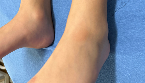
Image 2. Click to enlarge.

Osteomyelitis is one of the most prevalent musculoskeletal infections plaguing pediatric populations.1 Pathogenically it can develop by hematologic spread or via direct inoculation with significant penetrating trauma or surgery.1 The most common culprit is Staphylococcus aureus, as in our case. Presentation of osteomyelitis is varied and dependent on the extent of the infection. On one end of the spectrum, it can be a localized infection present on a single metaphysis free of associated symptoms. The other extreme is severe multifocal infection accompanied by septic shock.2

Image 3. Click to enlarge.

Our patient was misdiagnosed initially but, fortunately, a positive blood culture helped identify the diagnosis, and the patient was treated appropriately prior to development of permanent sequelae. The diagnosis of osteomyelitis is often difficult as it presents subclinically in the early stages and is relatively rare. Fever and pain are the most common presenting symptoms of osteomyelitis and can manifest as difficulty bearing weight or reduced use of the affected limb in pediatric patients.2 In our case the patient did have fever and a limp which was presumed secondary to sprain from the preceding dance lesson. A minimal skin abnormality consistent with inflammation was present, likely indicating the patient had an indolent infection. The use of inflammatory markers can guide the treatment plan, and an X-ray might show evidence of osteomyelitis. 3 Those were inconclusive on our patient and an MRI on initial presentation would have been ideal for diagnosis.3

Teaching Points

  • Unresolved fever in a pediatric patient should prompt further workup for osteomyelitis, especially if they have diabetes.
  • Inflammatory markers and X-ray imaging can be negative early in the disease course.
  • A skin rash may indicate infection in deeper tissue.

The prevalence of osteomyelitis in children within developed nations is 13 per 100,000, however it is higher among those with diabetes.4,5 Research evaluating the intersectionality of type 1 diabetes mellitus and osteomyelitis in pediatric patients is extremely limited, therefore calculating an exact prevalence rate is difficult. It has been determined that patients with diabetes have a 1.5-4.5 times greater risk of infection than the general population.5 This makes the diagnosis a pertinent consideration when a diabetic patient presents with fever from an unclear source.


Dr. Khalil Mroue is a clinical assistant professor at Burrell College of Osteopathic Medicine and at Hyatt Clinical Education. He currently works in emergency medicine at the Tucson Medical Center.

Dominic Barandica is a fourth-year medical student at the Autonomous University of Guadalajara.


References

  1. Walter N, Bärtl S, Alt V, Rupp M. The epidemiology of osteomyelitis in children. Children (Basel). 2021;8(11):1000. doi:10.3390/children8111000.
  2. Woods CR, Bradley JS, Chatterjee A, et al. Clinical practice guideline by the Pediatric Infectious Diseases Society and the Infectious Diseases Society of America: 2021 guideline on diagnosis and management of acute hematogenous osteomyelitis in pediatrics. J Pediatric Infect Dis Soc. 2021;10(8):801-844. doi:10.1093/jpids/piab027.
  3. Hackenberg RK, Schmitt-Sánchez F, Endler C, et al. Value of Diagnostic Tools in the Diagnosis of Osteomyelitis: Pilot Study to Establish an Osteomyelitis Score. J Clin Med. 2023;12(9):3057. Published April 23, 2023.. doi:10.3390/jcm12093057.
  4. Momodu II, Savaliya V. Osteomyelitis. In: StatPearls. StatPearls Publishing; 2023. Accessed September 19, 2025. https://www.ncbi.nlm.nih.gov/books/NBK532250/.
  5. Holt RIG, Cockram CS, Ma RCW, Luk AOY. Diabetes and infection: Review of the epidemiology, mechanisms and principles of treatment. Diabetologia. 2024;67:1168-1180. doi:10.1007/s00125-024-06102-x.

Topics: FeverInfectious DiseaseOsteomyelitisPediatricsStaphylococcus Aureustype 1 diabetes

Related

  • FACEPs in the Crowd: Dr. John Ludlow

    November 5, 2025 - 0 Comment
  • Nail Bed Injuries: What to Do—or Not to Do

    August 18, 2025 - 0 Comment
  • The Resurgence of Measles, Mumps, and Health Equity Challenges

    August 14, 2025 - 0 Comment

Current Issue

ACEP Now: December 2025 (Digital)

Read More

3 Responses to “Case Report: Five Days of Fever”

  1. December 21, 2025

    Gabe Wilson Reply

    A few learning points:

    1. The “Dissociated” Inflammatory Markers
    The most striking feature here was the discordance between the CRP (2.5 mg/L) and the Procalcitonin (2.24 ng/mL).
    • The Trap: Relying solely on the normal CRP led to the initial exclusion of osteomyelitis. In approximately 2–5% of AHO cases, the CRP can be normal on admission, particularly if the organism is less virulent (e.g., Kingella—though not the case here) or in early/walled-off S. aureus infections.
    • The Clue: The Procalcitonin was the “truth-teller.” A PCT >2.0 in a pediatric patient without a clear source is highly specific for invasive bacterial infection (bacteremia/sepsis) and should trigger a hunt for the source regardless of the CRP.
    2. The “Phantom” Limp
    The history of a limp that “improved” and a normal gait in the ED is a common confounder in pediatric osteomyelitis.
    • Children often guard intermittently.
    • The “lesion” on the ankle (initially thought to be the result of the dance shoe) was likely the cause (portal of entry) or a manifestation (septic embolus/Janeway lesion) of the intravascular infection.
    3. T1DM as a Risk Factor (co-morbidities, co-morbidities, co-morbidities – ALWAYS be super suspect of more advanced or unusual disease)
    Even with a normal glucose (81 mg/dL) and no DKA, the patient’s Type 1 Diabetes history is relevant.
    • It places the patient at higher risk for invasive S. aureus infections (via pump sites/CGM or altered neutrophil function).
    • It may also blunt the typical febrile/inflammatory response, contributing to the confusing presentation.

  2. December 21, 2025

    Gabe Wilson Reply

    One additional point:

    DIAGNOSTIC INCONGRUITY/ANCHORING BIAS

    I have yet to see such a subtle cellulitis in someone mounting a fever of 104F. With that fever, there would be a correspondingly large area of typical indurated, very erythematous, tender, warm skin.

    A subtle presentation on the derm exam combined with a fever of 104F does not, in my experience, align with a diagnosis of cellulitis.

  3. December 22, 2025

    STEPHEN J. VAN CLEAVE Reply

    Limping is not common with cellulitis. So with joint pain in a diabetic child, you need CT or MRI of the affected joint, not plain X-ray.

Leave a Reply Cancel Reply

Your email address will not be published. Required fields are marked *


*
*


Wiley
  • Home
  • About Us
  • Contact Us
  • Privacy
  • Terms of Use
  • Advertise
  • Cookie Preferences
Copyright © 2026 by John Wiley & Sons, Inc. All rights reserved, including rights for text and data mining and training of artificial technologies or similar technologies. ISSN 2333-2603