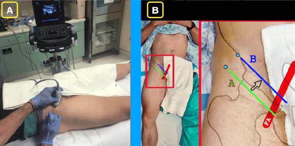
Figure 2A: The ultrasound system is placed contralateral to the affected hip so that the operator has a clear line of sight of the needle as well as the ultrasound screen.
Figure 2B: Basic position of the ultrasound transducer in the region of the inguinal ligament. The green line (A) indicates the first position. In this position, the femoral head and femoral artery will be located. The blue line (B) indicates a minor adjustment in order to locate the ideal location for the PENG block.

No Responses to “Figure 2A: The ultrasound system is placed contralateral to the affected hip so that the operator has a clear line of sight of the needle as well as the ultrasound screen. Figure 2B: Basic position of the ultrasound transducer in the region of the inguinal ligament. The green line (A) indicates the first position. In this position, the femoral head and femoral artery will be located. The blue line (B) indicates a minor adjustment in order to locate the ideal location for the PENG block.”