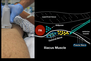
Figure 6: When performing an in-plane lateral to medial approach to the ultrasound-guided femoral nerve block, obtain clear sonoanatomy by proper probe positioning. Try to get your block needle just under the fascia iliaca (lateral to the femoral nerve) and inject anesthetic gently.

Figure 6: When performing an in-plane lateral to medial approach to the ultrasound-guided femoral nerve block, obtain clear sonoanatomy by proper probe positioning. Try to get your block needle just under the fascia iliaca (lateral to the femoral nerve) and inject anesthetic gently.

No Responses to “Figure 6: When performing an in-plane lateral to medial approach to the ultrasound-guided femoral nerve block, obtain clear sonoanatomy by proper probe positioning. Try to get your block needle just under the fascia iliaca (lateral to the femoral nerve) and inject anesthetic gently.”