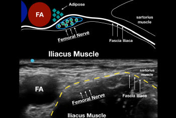
Figure 1: A. Drawing of the relevant sonoanatomy when performing an ultrasound-guided femoral nerve block. Note that the fascia iliaca keeps the femoral nerve right next to the iliacus muscle. B. Ultrasound image of the same anatomy. The fascia iliaca (yellow dotted line) is the key structure to recognize when performing the block.

Figure 1: A. Drawing of the relevant sonoanatomy when performing an ultrasound-guided femoral nerve block. Note that the fascia iliaca keeps the femoral nerve right next to the iliacus muscle. B. Ultrasound image of the same anatomy. The fascia iliaca (yellow dotted line) is the key structure to recognize when performing the block.

No Responses to “Figure 1: A. Drawing of the relevant sonoanatomy when performing an ultrasound-guided femoral nerve block. Note that the fascia iliaca keeps the femoral nerve right next to the iliacus muscle. B. Ultrasound image of the same anatomy. The fascia iliaca (yellow dotted line) is the key structure to recognize when performing the block.”