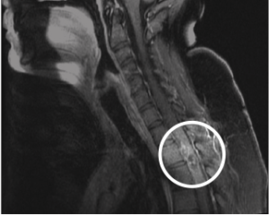
Figure 2: Thoracic MRI showing areas of hemorrhage in the spinal cord most prominent at the T2–T3 level, with multiple serpentine structures, most in keeping with a spinal cord vascular malformation such as an sAVM.
Figure 2: Thoracic MRI showing areas of hemorrhage in the spinal cord most prominent at the T2–T3 level, with multiple serpentine structures, most in keeping with a spinal cord vascular malformation such as an sAVM.
By Joseph Harrington
|
on December 15, 2020
|
0 Comment


No Responses to “Figure 2: Thoracic MRI showing areas of hemorrhage in the spinal cord most prominent at the T2–T3 level, with multiple serpentine structures, most in keeping with a spinal cord vascular malformation such as an sAVM.”